

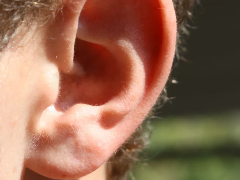

For decades, a pervasive biological myth claimed that human ears and noses possess a mysterious superpower of limitless lifelong growth. However, rigorous anatomical research has conclusively settled the debate: uncovering why do ears get bigger with age is not a story of eternal chondrocyte cell division, but rather an inevitable consequence of terrestrial gravity, collagen degradation, and the biomechanical breakdown of elastic connective tissues.
The Biomechanical Enigma: Why do ears get bigger with age and debunking endless growth
Examine vintage family portraits or observe the facial features of elderly relatives, and a striking anatomical pattern becomes impossible to overlook: older adults unquestionably possess longer, more prominent ears. In popular culture and everyday folklore, this observation fostered the stubborn belief that elastic cartilage is the only human tissue exempt from biological senescence, continuing to multiply and expand until our final breath.
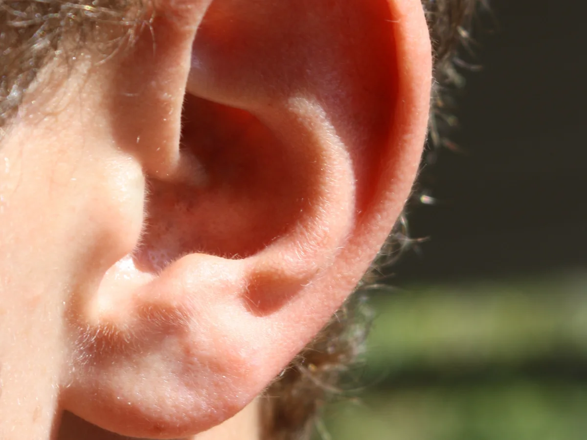
In reality, the proliferation of chondrocytes—the specialized cells responsible for producing cartilage—ceases entirely following puberty, coinciding with the fusion of skeletal growth plates. Throughout adult life, the auricular cartilage remains in a metabolic steady state characterized by virtually zero cellular replication. What visually masquerades as “growth” is fundamentally passive mechanical elongation driven by decades of gravitational pull and structural deterioration of dermal proteins.
The definitive clinical investigation into this phenomenon was pioneered by British physician James Heathcote (James Heathcote). In a landmark 1995 paper published in the British Medical Journal (BMJ), Dr. Heathcote measured the ear dimensions of 206 patients aged 30 to 93 years. His rigorous anthropometric analysis revealed that human ears lengthen by an average of 0.22 millimeters per year, accumulating more than 1.1 centimeters of downward stretch over a 50-year adult lifespan. Biomedical researchers investigating age-related structural changes often compare how the human body adapts over time, an inquiry closely tied to neurological and physical preservation explored in our analysis of cellular protein dynamics across the human lifespan.
Anatomy of the Auricle: Why the Earlobe Suffers the Greatest Deformation
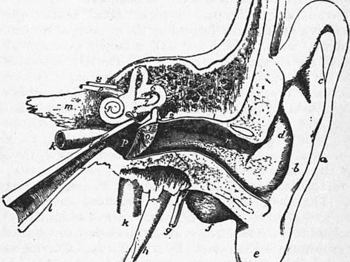
Unpacking the physical mechanics of ear elongation requires distinguishing between the two distinct anatomical regions of the external pinna: the upper two-thirds, supported by a semi-rigid plate of elastic cartilage, and the inferior third—the earlobe (lobulus auriculae). Unlike the helix and antihelix, the earlobe contains absolutely no cartilage scaffolding. It consists entirely of adipose tissue, loose vascularized stroma, and an outer cutaneous envelope.
With progressive chronological aging, the dermis undergoes a catastrophic reduction in two structural fibrillar proteins: collagen types I and III, which provide tensile strength, and elastin, which enables tissues to recoil after mechanical deformation. Dermal fibroblast synthesis drops by approximately 1% each year after age 25, while environmental factors such as ultraviolet solar radiation (solar elastosis) enzymatically degrade existing protein cross-links.
Deprived of its internal elastic resilience, the fleshy lobule gradually yields to its own weight and the relentless downward acceleration of gravity. In women, this elongation is substantially exacerbated by decades of wearing heavy earrings, where chronic static tensile load produces permanent plastic creep of the connective tissue, resulting in dramatically stretched earlobes.
«Human ears undeniably elongate with age at a rate of roughly 0.22 millimeters per year. This is not biological growth, but rather the inexorable consequence of gravity pulling down on declining elastic tissues.»
Dr. James Heathcote (James Heathcote), British General Practitioner and Fellow of the Royal College of General Practitioners
Degradation of Elastic Cartilage: Glycation, Calcification, and Matrix Collapse
Even though the upper auricle is braced by cartilage, it is far from immune to age-related metamorphosis. Auricular elastic cartilage is permeated by an intricate three-dimensional network of elastin fibers that, in youth, grant the ear its spring-like recoil. Over decades, non-enzymatic glycation—the covalent bonding of glucose molecules to proteins—induces pathological cross-linking, causing elastin fibers to stiffen, micro-calcify, and shatter under microtrauma.
Concurrently, the extracellular matrix loses its water-binding proteoglycans and hyaluronic acid polymers. This osmotic dehydration reduces internal hydrostatic turgor, causing the cartilage framework to thin out and flatten. As the three-dimensional helical folds uncurl, the pinna widens and flattens against the temporal plane, visibly expanding its projected surface area when viewed from the front.
Forensic anthropologists at the University of Milan (University of Milan), studying anthropometric ear databases of more than 1,400 individuals, established that pinna dimensional ratios serve as one of the most reliable biometric markers for biological age estimation. Male ears average significantly larger dimensions than female ears across all adult age brackets, driven by larger baseline craniofacial proportions and lifelong androgenic influences on connective tissue thickness.
The documented average rate of adult ear elongation identified in landmark British Medical Journal anthropometric trials.
Adult chondrocytes do not replicate; ears lengthen through gravitational stretching and matrix creep rather than new tissue growth.
An annual 1% decline in dermal collagen synthesis strips the lobule of its structural tensile strength and elasticity.
Pinna uncurling and elongation alters acoustic resonance between 2–5 kHz, influencing vertical sound localization in older adults.
The Craniofacial Optical Illusion: Gravitational Ptosis and Bone Resorption
The visual perception of elderly ears as disproportionately enormous is magnified by concurrent atrophy of the surrounding facial skeleton. Normal facial aging is characterized by deflation and descent of deep subcutaneous fat compartments across the cheeks, temples, and malar prominence. Soft tissues migrate downward under gravitational ptosis, while the underlying maxilla and mandible undergo progressive osteoclast-mediated bone resorption, particularly following tooth loss.
Consequently, the middle and lower thirds of the human face lose volume and retract inward. Against this diminishing, gaunt facial canvas, ear auricles that have elongated by just 8 to 12 millimeters over a lifetime appear visually dominant and oversized. Human visual cognition evaluates feature proportions comparatively rather than against absolute scale measurements.
Beyond aesthetics, structural changes to the pinna alter auditory psychoacoustics. The intricate convolutions of the concha and helix act as a directional resonant horn, shaping the frequency spectrum between 2 and 5 kHz to aid horizontal and vertical sound localization. As the auricle flattens and droops, its acoustic transfer function shifts, which compounds presbycusis (age-related sensory hearing loss) and prompts older individuals to cup their hand behind the ear to manually restore acoustic resonance.
- Ears do not grow endlessly: active chondrocyte cell division halts permanently upon reaching adult maturity.
- The earlobe contains no cartilage and is composed strictly of fat and skin, making it highly susceptible to gravitational sagging.
- Solar ultraviolet exposure degrades dermal elastin fibers, accelerating irreversible structural drooping.
- Age-related bone and fat loss in the cheeks and jaw shrinks facial volume, creating an optical illusion of oversized ears.