

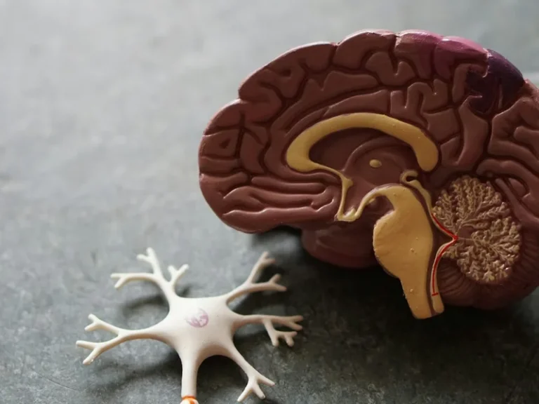

Traversing the deepest conduits of human anatomy lies the most extensive and influential conduit of the parasympathetic nervous system: the tenth cranial nerve, canonically designated the vagus nerve (Nervus Vagus). Originating within the medullary nuclei of the brainstem, this neural trunk arborizes across the cervical region, heart, pulmonary tree, and gastrointestinal viscera, constituting an unbroken bidirectional superhighway between mind and body. Groundbreaking neuroimmunological research has established that the vagus nerve vagal tone heart rate variability inflammation serves as the master homeostatic switchboard of human physiology, governing the cholinergic anti-inflammatory pathway via alpha-7 nicotinic acetylcholine receptors (alpha-7 nAChR). Targeted vagal modulation via transcutaneous auricular stimulation (taVNS) and resonance frequency breathing protocols can extinguish inflammatory cytokine storms, optimize sleep architecture, and dissolve the debilitating burden of chronic autonomic stress.
Anatomy of the Tenth Cranial Nerve: The Autonomic Superhighway
The vagus nerve constitutes the tenth and by far the longest of the twelve cranial nerve pairs. Emerging from paired motor and sensory nuclei within the medulla oblongata—specifically the dorsal motor nucleus, nucleus ambiguus, and nucleus tractus solitarii—the vagal trunks exit the cranial vault through the jugular foramen. Descending through the carotid sheath between the internal carotid artery and internal jugular vein, the vagus arborizes extensively across the pharynx, larynx, heart, trachea, bronchi, esophagus, stomach, liver, pancreas, spleen, and intestinal tract down to the splenic flexure.
A fundamental organizational feature of the vagus is its fiber distribution: more than 80% of all axons are afferent sensory fibers. Rather than carrying motor commands from the central nervous system, these sensory highways continuously funnel a colossal volume of interceptive feedback from peripheral organs back into the brainstem. The vagus serves as the primary data conduit of the gut-brain axis, updating the brain regarding microbiome metabolic byproducts, luminal osmolarity, mucosal cytokines, and mechanical visceral distension.
The remaining efferent fibers represent preganglionic parasympathetic motors. When stimulated, terminal boutons release acetylcholine into cardiac synapses, engaging muscarinic M2 receptors across the sinoatrial and atrioventricular nodes. This exerts a rapid negative chronotropic effect, dampening heart rate and restoring systemic hemodynamic calm.

The Cholinergic Anti-Inflammatory Pathway: Kevin Tracey’s Discovery
For generations, immunology and neuroscience operated as separate disciplines under the belief that systemic immune defenses operated autonomously via circulating humoral mediators. This dogma was dismantled in the early 2000s when pioneering neurosurgeon Kevin Tracey uncovered the cholinergic anti-inflammatory pathway.
Tracey discovered that upon detecting peripheral inflammatory cascades, the brain mobilizes the efferent vagus nerve as an instantaneous neurological circuit breaker. Vagal action potentials travel down to the celiac-superior mesenteric ganglion complex, relaying signals along the splenic nerve into the spleen—the primary reservoir of systemic immune cells. Within the splenic parenchyma, nerve fibers interface with a specialized subset of acetylcholine-synthesizing T cells (ChAT+ T cells).
These specialized lymphocytes release acetylcholine, which selectively binds to alpha-7 nicotinic acetylcholine receptors (alpha-7 nAChR) expressed on the surface of splenic macrophages. This receptor engagement inhibits the nuclear translocation of NF-kappaB, shutting down transcription of catastrophic pro-inflammatory cytokines including tumor necrosis factor-alpha (TNF-alpha), interleukin-1-beta (IL-1beta), and interleukin-6 (IL-6). Robust vagal tone thus shields the organism from systemic low-grade inflammation, septic shock, rheumatoid arthritis, and atherosclerosis.
Efferent vagal acetylcholine binds to alpha-7 nicotinic acetylcholine receptors on splenic macrophages, suppressing TNF-alpha, IL-1beta, and IL-6 synthesis.
The millisecond beat-to-beat variability of consecutive R-R intervals represents the gold-standard biomarker of vagal parasympathetic tone and physiological resilience.
Non-invasive bioelectrical stimulation of the auricular vagus branch at the tragus directly modulates brainstem autonomic nuclei, enhancing slow-wave restorative sleep.
Over 80% of vagal fibers are sensory afferents, transmitting continuous real-time biochemical telemetry regarding the microbiome and enteric inflammatory state to the brain.
Heart Rate Variability (HRV) as the Mirror of Parasympathetic Health
Assessing vagal nerve function non-invasively represented a major clinical dilemma until cardiologists and neurophysiologists recognized its mathematical signature within Heart Rate Variability (HRV).
A healthy human heart does not beat with the rigid cadence of a metronome. At a resting pulse of 60 beats per minute, the intervals separating consecutive R-waves on an electrocardiogram vary continuously: 1,020 ms, 940 ms, 1,060 ms. This dynamic irregularity reflects the constant tug-of-war between the sympathetic accelerator and the parasympathetic vagal brake.
During inspiration, medullary sympathetic centers transiently inhibit the vagus, accelerating heart rate; upon expiration, the vagus re-engages instantaneously, releasing acetylcholine to prolong inter-beat intervals (respiratory sinus arrhythmia). Spectral power analysis of the high-frequency band (HF, 0.15–0.40 Hz) and the root mean square of successive differences (RMSSD) provide high-precision proxies of vagal tone. High HRV correlates with cardiovascular longevity, neuroplasticity, and emotional stability, whereas depressed HRV is a potent harbinger of cardiac mortality and depression.
«The vagus nerve functions as our physiological braking system. When sympathetic stress depresses the accelerator, vagal tone protects vital organs from burnout, oxidative stress, and chronic inflammation.»
Dr. Stephen Porges, Distinguished University Scientist and Originator of the Polyvagal Theory
Transcutaneous Auricular Stimulation (taVNS): Non-Invasive Neuromodulation
Recognizing the transformative therapeutic impact of vagal activation led to surgically implantable vagus nerve stimulators (VNS) wrapped around the cervical trunk for drug-resistant epilepsy and refractory depression. However, consumer medicine achieved a breakthrough with transcutaneous auricular vagus nerve stimulation (taVNS).
The anatomical basis of taVNS relies on a unique evolutionary trait: the external ear is the sole surface on the human body where sensory vagal fibers terminate directly beneath the cutaneous epidermis. The auricular branch (ramus auricularis, or Arnold’s nerve) innervates the tragus, cymba conchae, and acoustic meatus.
Applying lightweight surface clips with medical-grade titanium electrodes to the tragus delivers mild, imperceptible bioelectrical pulses at 20–30 Hz. Ascending afferents carry the stimulus into the nucleus tractus solitarii, which in turn modulates the locus coeruleus (noradrenaline) and dorsal raphe nuclei (serotonin). Rigorous clinical trials confirm that daily 20-minute taVNS protocols recalibrate autonomic balance, attenuate migraine intensity, improve slow-wave sleep depth, and lower circulating C-reactive protein without pharmaceutical side effects.
- The Latin designation «Vagus» translates to ‘wandering’, describing its vast trajectory from brainstem to colon.
- Resonance breathing at 5.5 to 6 breaths per minute maximizes respiratory sinus arrhythmia and arterial baroreflex sensitivity.
- Invasive vagus nerve stimulation is clinically approved for refractory major depression and intractable epilepsy.
- Cold face immersion triggers the mammalian dive reflex, instantaneously inducing vagal bradycardia.
Natural Activation Protocols: Resonance Frequency Breathing and Cold Shock
Beyond electronic therapeutics, human physiology possesses innate behavioral levers for stimulating vagal tone, readily deployable in daily life. Foremost among these is Resonance Frequency Breathing.
Slowing respiration to a paced rate of approximately 5.5 to 6 breaths per minute (inhaling smoothly for 4 seconds and exhaling unhurriedly through the nose for 6 seconds) harmonizes respiratory rhythm with endogenous 0.1 Hz Mayer waves of blood pressure regulation. This mechanical resonance amplifies baroreflex sensitivity, triggering a massive parasympathetic rebound: blood pressure stabilizes, cerebral blood flow increases, and acute anxiety dissipates within minutes.
A second powerful physiological activator is the mammalian dive reflex. Submerging the face in cold water (10–12 degrees Celsius) or placing a chilled compress over the neck stimulates ophthalmic trigeminal and cervical vagal fibers, triggering instant reflex bradycardia and peripheral vasoconstriction. Cultivating vagal tone represents an empirical, science-backed foundation for long-term physiological resilience, cognitive clarity, and vitality. Also read our related investigative feature on evidence-based physiological methods for autonomic nervous system regulation and stress reduction.