

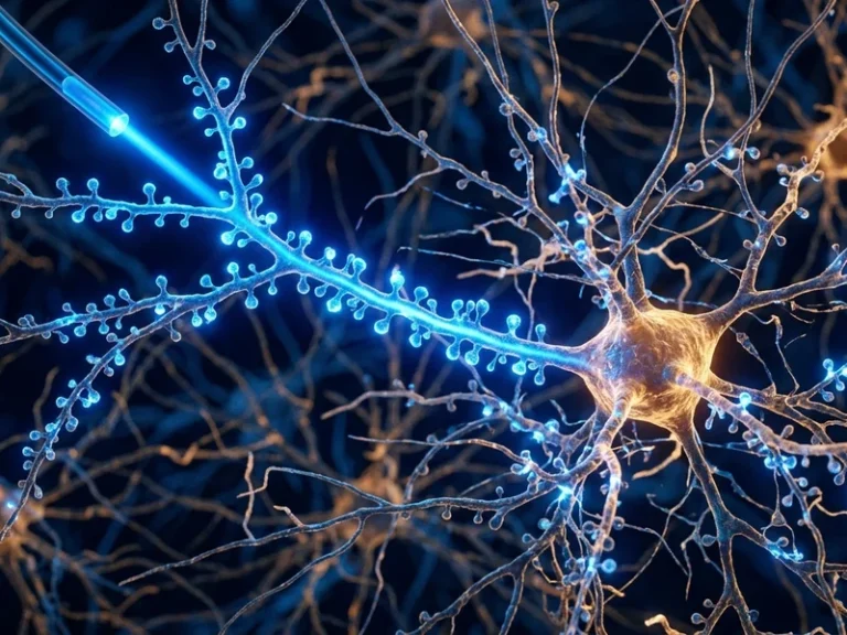

When researchers in the early 2000s first attempted to express light-sensitive microbial proteins from unicellular algae inside mammalian neurons, mainstream neuroscience viewed the concept with profound skepticism. Today, however, optogenetics neural control parkinsons disease brain has transformed from an eccentric bioengineering experiment into one of the most revolutionary clinical and investigative paradigms of modern medicine. By delivering targeted laser pulses through microscopic optical fibers into deep cerebral architectures, scientists can selectively activate or silence genetically designated neuronal ensembles with millisecond precision, illuminating unprecedented avenues for curing neurodegenerative disorders, blindness, and treatment-resistant depression.
The Dawn of a Paradigm: From Algal Photosensing to Mammalian Neurons
The origins of optogenetics trace back to fundamental biophysical investigations of the green unicellular alga Chlamydomonas reinhardtii. In the late 1990s and early 2000s, German biophysicists Peter Hegemann and Georg Nagel isolated and characterized Channelrhodopsin-1 and Channelrhodopsin-2. These extraordinary transmembrane proteins fused the functionality of a light sensor directly to an ion channel: upon absorbing a blue photon with a wavelength of approximately 470 nanometers, the pore opened, precipitating a rapid influx of sodium and calcium cations into the cytosol.
The conceptual leap that transformed biology occurred at Stanford University in 2005, spearheaded by Karl Deisseroth alongside graduate students Edward Boyden and Feng Zhang. They packaged the algal gene into a benign adeno-associated viral (AAV) vector and introduced it into mammalian cortical neurons. The experiment proved an instant sensation: when illuminated by blue light pulses, the engineered neurons generated authentic action potentials within milliseconds, establishing precise optical neuromodulation.
Soon after, researchers identified Halorhodopsin from the haloarchaeon Natronomonas pharaonis, which pumped chloride anions into the cell under amber-yellow light, hyperpolarizing the membrane and instantly quenching neural firing. With this discovery, neuroscience acquired a true binary switchboard: blue light switches neurons on, while yellow light switches them off, bypassing the imprecise chaos of electrical electrodes.
Crystallographic elucidations of channelrhodopsin chimeras revealed the atomic precision of light-induced pore opening. The retinal chromophore, covalently anchored to a conserved lysine residue via a protonated Schiff base, undergoes ultrafast trans-to-cis photoisomerization within 200 femtoseconds of photon absorption. This triggers tilt motions across the transmembrane alpha-helices, forming a hydrated central cation pathway conducting sodium and calcium currents.
Engineered opsin variants such as Chronos and ChETA broke the temporal speed limit of optical neuromodulation. While wild-type ChR2 exhibited channel closure deactivation kinetics of 10-15 milliseconds—restricting spike fidelity above 40 Hz—modern synthetic opsins follow firing rates exceeding 200 Hz, faithfully mirroring the physiological firing dynamics of parvalbumin-positive interneurons.
Conquering Parkinson’s: Deconstructing Basal Ganglia Circuitry
One of optogenetics’ most profound clinical triumphs has been deciphering the enigmatic microcircuit pathology of Parkinson’s disease. Historically, the disease was viewed primarily as the inexorable loss of dopaminergic neurons in the substantia nigra pars compacta. While deep brain stimulation (DBS) provided symptomatic relief, its underlying mechanism remained controversial because electrical currents indiscriminately stimulated every axon and soma within the subthalamic nucleus.
Employing cell-type-specific optogenetics in rodent and non-human primate models, Deisseroth’s group uncovered an astonishing reality: the decisive therapeutic factor in alleviating motor symptoms is not the inhibition of subthalamic neurons themselves, but the selective activation of afferent axonal projections originating in the primary motor cortex. Illuminating these specific pathways instantly abolished bradykinesia and motor tremors within fractions of a second.
These insights have inspired closed-loop adaptive DBS architectures. By sensing pathological beta-band oscillations in real time and delivering calibrated neurostimulation exclusively during motor failure episodes, next-generation implants minimize battery drain and avoid disruptive cognitive side effects.
Pathophysiologically, the hallmark motor deficits of Parkinson’s disease arise from hyper-synchronized beta-band oscillations (13-30 Hz) throughout the subthalamic nucleus and globus pallidus. Conventional electrical DBS suppresses this pathological rhythm via non-specific current dispersal, frequently triggering collateral affective and speech impairments.
Targeted optogenetic interrogation revealed that activating specific afferent axonal projections descending from the motor cortex fully rescues motor initiation and extinguishes tremor. This paradigm underpins the engineering of closed-loop adaptive optoelectronic neuroprosthetics that optically damp pathological synchronization only when aberrant bursts emerge.
Optogenetic switches fire within 1-2 milliseconds, matching the authentic physiological speed of endogenous action potentials.
Blue light activates neurons via Channelrhodopsin, while amber-yellow light instantly suppresses neural firing via Halorhodopsin.
Opsins convert surviving ganglion cells into artificial photoreceptors, restoring functional vision in blind patients.
Unlike electrical stimulation, light exclusively manipulates genetically specified cell types, avoiding collateral damage.
Restoring Vision: Converting Surviving Retinal Cells into Photoreceptors
Ophthalmology has become the first discipline where optogenetic gene therapy has crossed the chasm into human clinical efficacy. Inherited diseases such as retinitis pigmentosa and advanced macular degeneration lead to the devastating destruction of rod and cone photoreceptors, plunging millions into total darkness. Crucially, however, the inner retinal layers—including bipolar cells and retinal ganglion cells—often remain viable and structurally wired to the visual cortex.
In 2021, a landmark clinical trial led by Dr. José-Alain Sahel and published in Nature Medicine reported that a 58-year-old completely blind patient had partially regained functional vision. Clinicians injected an adeno-associated virus encoding the red-shifted channelrhodopsin ChrimsonR into his eye, transforming surviving ganglion cells into light-sensitive units.
Coupled with biometric neuromorphic goggles that translated ambient optical scenes into targeted amber-light pulses, the patient was able to perceive high-contrast objects on a table, count items, and navigate crosswalks. This demonstrated for the first time that optogenetic cellular reprogramming can restore functional sensory perception in humans.
The clinical translation of vision restoration via ChrimsonR highlights the vital role of photon density engineering. Endogenous photoreceptors possess vast biochemical amplification cascades mediated by transducin and cGMP-phosphodiesterase, enabling single-photon detection. Microbial opsins function as direct passive ion gates, demanding substantial photonic flux.
Specialized bioengineered goggles overcome this thermodynamic deficit by capturing scene dynamics with event-based neuromorphic sensors and projecting amber-spectrum pulses (595 nm) onto transduced ganglion cells, driving perception without causing photothermal damage to the delicate retinal pigment epithelium.
«Optogenetics has fundamentally dismantled the wall between passively observing brain activity and actively steering it. We can now read, dissect, and rewrite the neural code in real time.»
Karl Deisseroth, Professor of Bioengineering and Psychiatry at Stanford University
Remapping the Depressive Circuit: Anhedonia and PTSD Ensembles
Major depressive disorder and post-traumatic stress disorder (PTSD) have long confounded conventional psychopharmacology, with over thirty percent of patients failing to respond to standard monoaminergic drugs. Neuroimaging reveals that depression is fundamentally a disorder of distributed brain circuit synchronization across the prefrontal cortex, amygdala, and nucleus accumbens rather than a simplistic chemical deficit.
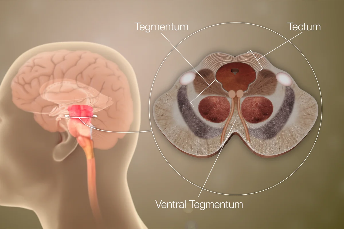
Through optogenetic dissection, neurobiologists mapped the specific circuitry governing anhedonia—the hallmark inability to experience pleasure. By selectively activating dopaminergic projections from the ventral tegmental area to the nucleus accumbens, researchers successfully reversed profound despair and chronic stress phenotypes in animal models within minutes.
In PTSD research, scientists have successfully tagged fear memory engram cells within the hippocampus with light-gated opsins. By applying low-frequency optical silencing to these specific neural traces, researchers neutralized traumatic fear associations without degrading neutral autobiographical memories.
In neuropsychiatry, optogenetics systematically dismantled the monolithic dopamine hypothesis. Precise circuit mapping demonstrated that distinct dopaminergic pathways originating in the ventral tegmental area serve orthogonal behavioral roles: projections to the medial prefrontal cortex mediate aversive stress coping, whereas projections to the nucleus accumbens govern motivational salience and hedonic drive.
Phase-locked 20-Hz photostimulation of the mesolimbic circuit immediately reverses chronic stress-induced anhedonia, validating network-level dysregulation as the biological substrate of mood disorders.
- 2002-2003: Peter Hegemann and Georg Nagel discover Channelrhodopsin light-gated ion channels.
- 2005: Karl Deisseroth at Stanford demonstrates the first optical control of mammalian neurons.
- 2021: First successful partial restoration of vision in a blind retinitis pigmentosa patient by Sahel et al.
- 2025-2026: Emergence of near-infrared upconversion nanoparticles for completely non-invasive deep-brain neuromodulation.
The Non-Invasive Frontier: Upconversion Nanoparticles and Future Horizons
Despite its breathtaking capabilities, clinical adoption of optogenetics has been restricted by the requirement for invasive intracerebral fiber-optic implants. However, the confluence of nanotechnology and photonics is overcoming this hurdle through upconversion nanoparticles (UCNPs).
UCNPs act as local nanoscale photon converters: they absorb deep-penetrating, near-infrared (NIR) light that travels harmlessly through the skull and scalp, and upconvert it into localized blue or green photons directly adjacent to opsin-expressing neurons, enabling completely non-invasive deep-brain optical neuromodulation.
As optogenetics converges with CRISPR-guided genomic targeting and wireless microelectronics, the future of medicine is entering an era where psychiatric afflictions, chronic neuropathic pain, and sensory impairments will be remediated not with indiscriminate chemicals, but with the surgical precision of light.
Next-generation biophotonics emphasizes transient, bioresorbable waveguides synthesized from silk fibroin and polylactic acid (PLA). These bioresorbable conduits deliver therapeutic illumination to deep brain targets and systematically dissolve into benign metabolites, avoiding chronic neuroinflammatory glial scarring.
Coupled with rare-earth upconversion nanoparticles that convert transcranial near-infrared rays into localized visible spectra, optogenetics is crossing the threshold into non-invasive clinical reality. Also read our related investigative feature on evolutionary mechanisms of nervous system architecture.