

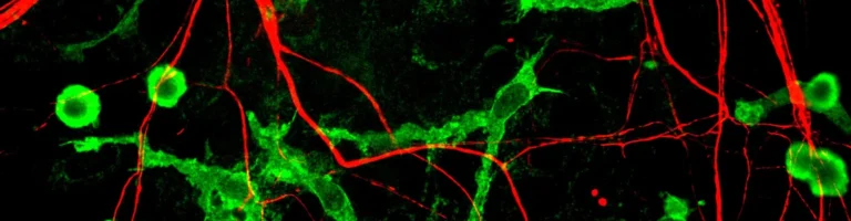

Every single night, as human consciousness transitions into slow-wave non-REM sleep, an intricate cellular renovation unfolds throughout the biological architecture of our brain. The principal conductors of this restorative maintenance are microglia — the resident immune sentinels of the central nervous system, which morph into meticulous gardeners of our neural circuits under the cover of darkness. Guided by the classical complement cascade, these ramified cells tirelessly interrogate billions of synaptic contacts, tagging underutilized dendritic spines and phagocytosing them via targeted trogocytosis. Without this vital nocturnal restructuring, the mammalian brain would swiftly collapse under informational static, cognitive fatigue, and neurodegenerative decline.
Resident Sentinels of the CNS: From Immune Defense to Synaptic Refinement
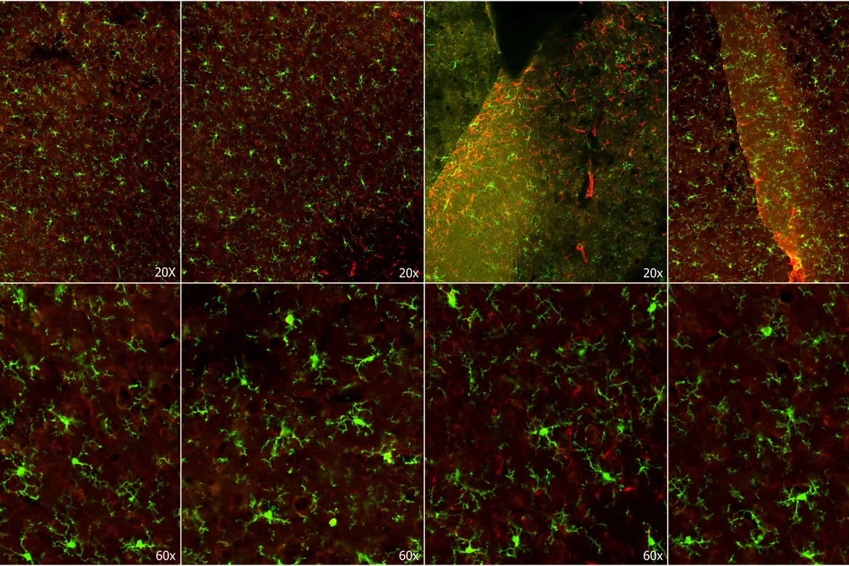
For decades, traditional neuroscience viewed microglia merely as passive scavengers — phagocytic cleanup cells mobilized only in response to trauma, bacterial infection, or ischemic stroke. However, modern high-resolution two-photon in vivo imaging has revealed that in the healthy, uninjured brain, microglia serve as dynamic architects of synaptic plasticity.
Microglial cells continuously scan their tissue microenvironment using exceptionally motile, branch-like cytoplasmic extensions. Within any 24-hour cycle, the processes of an individual microglial cell make transient, direct physical contact with thousands of presynaptic and postsynaptic boutons, evaluating their molecular and electrical integrity.
During wakefulness, the ceaseless flood of environmental stimuli drives widespread synaptogenesis. Left unchecked, this uncontrolled proliferation would overwhelm the brain metabolic resources and trigger catastrophic circuit hyper-synchrony.
The Tagging Cascade: Classical Complement Factors C1q and C3 as Removal Beacons
How does a microglial cell discern an essential memory trace from an obsolete synaptic junction? The underlying mechanism relies on the classical complement cascade, co-opted from innate immunity by the central nervous system.
When a synaptic connection is seldom depolarized and its physiological activity wanes, its dendritic membrane exhibits localized molecular cues that bind the initiator protein C1q. This binding triggers a downstream enzymatic cascade that deposits complement fragment C3 — an unmistakable molecular «eat-me» tag — onto the doomed synapse.
Microglial processes abundantly express Complement Receptor 3 (CR3). Upon binding C3, the microglial cell initiates trogocytosis — physically nibbling and internalizing the tagged dendritic spine, directing it to acidic lysosomal compartments for enzymatic degradation while preserving the parent neuronal axon.
Beyond the classical C1q and C3 cascade, recent investigations have revealed the pivotal role of externalized phosphatidylserine. In silent dendritic spines, this phospholipid flips to the outer membrane leaflet, serving as an unambiguous conformational trigger for microglial TREM2 and GPR56 receptors, guaranteeing strict biochemical fidelity before spine internalization.
Innate complement proteins mark dormant dendritic spines with an eat-me tag for targeted microglial phagocytosis.
Nocturnal decrease in norepinephrine removes inhibition, greatly accelerating microglial branch motility and engulfment.
Slow-wave sleep prunes informational noise, restoring metabolic equilibrium and consolidating vital memory engrams.
Prolonged sleep debt shifts microglia into an aberrant state where they consume healthy synapses and myelin.
Why Pruning Occurs During Sleep: The Synaptic Homeostasis Hypothesis
The Synaptic Homeostasis Hypothesis (SHY), pioneered by Giulio Tononi and Chiara Cirelli at the University of Wisconsin–Madison, posits that wakefulness causes a net net increase in synaptic weight across the cortex, which is unsustainable in terms of space and cellular energy.
During sleep, brain levels of vigilance-promoting neuromodulators — particularly norepinephrine, serotonin, and histamine — decrease sharply. The drop in ambient norepinephrine acts as a permissive chemical signal for microglia: their ramified processes double their motility and phagocytic appetite.
Unburdened by sensory processing, the brain enters synchronized slow-wave sleep. In this phase, heavily consolidated engrams express protective «don’t-eat-me» molecular shields (such as CD47), while noisy background connections are systematically cleared away.
«Microglia during sleep are by no means dormant observers waiting for pathogen invasion. Instead, they dynamically survey cortex and hippocampus, physically eliminating redundant synapses. Sleep represents a crucial physiological window where the brain resets its computational hardware.»
Beth Stevens, Associate Professor of Neurology at Harvard Medical School and Boston Children’s Hospital
Chronic Sleep Deprivation and Neurodegeneration: When Sentinels Consume Healthy Neurons
What happens when sleep is chronically truncated? Circadian disruption shifts microglia from beneficial sculptors into an aberrant, hyper-reactive neuroinflammatory phenotype.
Pioneering work by Michele Bellesi at Marche Polytechnic University demonstrated that acute sleep loss prompts microglia to aggressively devour not only weak spines but also mature presynaptic terminals and myelin sheaths. The protective pruning turns into an uncontrolled autoimmune attack on intact neural networks.
Furthermore, defective pruning impairs the glymphatic clearance of neurotoxic oligomers, allowing beta-amyloid plaques and hyperphosphorylated tau to accumulate — the defining hallmarks of Alzheimer’s and Parkinson’s disease. Chronic insomnia literally traps cellular waste within the cerebral parenchyma.
- Microglia account for roughly 10–15% of all non-neuronal cells in the adult mammalian central nervous system.
- Selective synaptic pruning during slow-wave sleep is essential for Long-Term Potentiation (LTP) in the hippocampus.
- The CD47 membrane protein serves as an active «don’t-eat-me» shield, protecting robust memory engrams from removal.
- Defective synaptic pruning during critical developmental windows has been implicated in Autism Spectrum Disorders (ASD).
Actionable Insights for Cognitive Longevity: Supporting Nocturnal Brain Cleansing
Recognizing the role of microglial pruning underscores that sleep is not passive downtime, but an active metabolic reset. Sustaining optimal synaptic clearance requires adhering to fundamental chronobiological principles.
Maintaining 7 to 8 hours of uninterrupted nocturnal sleep in pitch-black darkness preserves deep slow-wave delta cycles when microglial remodeling peaks. Avoiding heavy caloric meals and stimulants 4 to 6 hours before sleep prevents peripheral inflammation from prematurely activating microglia.
Prioritizing healthy sleep hygiene safeguards cognitive sharpness, enhances memory consolidation, and builds a robust molecular barrier against age-related neurodegenerative decline. Читайте также наш связанный материал про neurobiological mechanisms of brain plasticity.