


A bizarre swallowed bone in the appendix, surgically retrieved by Italian doctors fifteen years after accidental ingestion, represents one of the most astonishing diagnostic dilemmas in contemporary gastrointestinal surgery.
A Bone in the Appendix: Diagnostic Enigma in the Emergency Room
When an adult male walked into an emergency department complaining of acute, localized pain in his lower-right abdomen, the attending surgical team immediately suspected a routine presentation. The discomfort centered on McBurney’s point, accompanied by focal tenderness and abdominal guarding, standard clinical hallmarks that typically indicate acute appendicitis. However, subsequent diagnostic testing left the clinical staff thoroughly astonished.
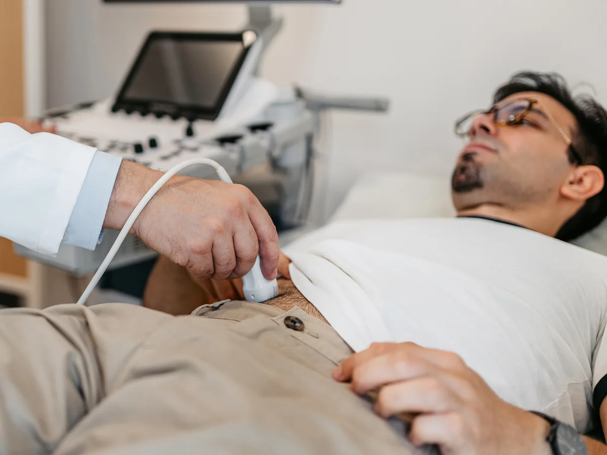
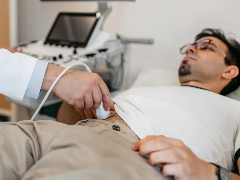
Despite sharp, recurrent pain, the patient showed zero signs of systemic inflammatory response. His body temperature was entirely normal, white blood cell counts were within standard physiological ranges, and markers of peritonitis or peritoneal irritation were completely absent. An urgent abdominal ultrasound scan revealed an unexpected result: the cecum, terminal ileum, and appendix itself appeared unremarkable, showing normal wall thickness, healthy luminal diameter, and no periappendiceal fluid accumulation.
Symptomatic treatment provided temporary pain relief, and the patient was discharged under watchful observation. Yet the underlying trigger persisted, hidden deep within the right iliac fossa for another eight months until debilitating abdominal cramps compelled him to seek emergency hospital care once again.
A Meal Forgotten for 15 Years: The Journey of an Ingested Bone
During his second hospitalization, surgeons conducted an extensive retrospective lifestyle interview. The patient suddenly recalled a distant, seemingly trivial memory: approximately 15 years earlier, during dinner, he had accidentally swallowed a long piece of bone containing edible marrow. Because he experienced no choking or esophageal pain at the time, he assumed stomach acids would dissolve the object and forgot about it entirely for a decade and a half.
To investigate the persistent pain, physicians performed an abdominal X-ray followed by an axial computed tomography (CT) scan with intravenous contrast. The imaging revealed a dense, well-circumscribed calcified mass measuring 2.5 by 3 centimeters lodged in the distal portion of the vermiform appendix.
Surgeons initiated a minimally invasive laparoscopic exploration and discovered an enlarged appendix containing an intraluminal hard mass. They performed a standard laparoscopic appendectomy. Upon dissecting the excised specimen, surgeons discovered a dense fecaloma (a hardened fecal stone) encasing an impeccably preserved piece of long bone. The clinical investigation was subsequently documented in the World Journal of Gastrointestinal Surgery.
Although the muscular walls of the appendix were distended by the growing mass, histological analysis confirmed an extraordinary finding: there were no cellular signs of active phlegmonous inflammation or necrosis. The patient recovered smoothly without complications and was discharged two days later.
«It is therefore possible, although rare, that a foreign body in the appendix even after many years (in our case, 15 years) does not lead to an authentic appendicitis either histologically or clinically.»
Nicolò Antonacci, MD, Department of General Surgery, Alma Mater Studiorum Università di Bologna
Mechanical Pathways: How Foreign Objects Enter the Vermiform Appendix
Annually in the United States alone, emergency medical services handle over 120,000 cases of ingested foreign objects, approximately 90% of which occur in children. In adults, common ingested items include chicken and fish bones, dental crowns, toothpicks, and small pieces of jewelry. More than 99% of smooth objects transit harmlessly through the gastrointestinal tract and are naturally expelled within 24 to 48 hours without medical intervention.
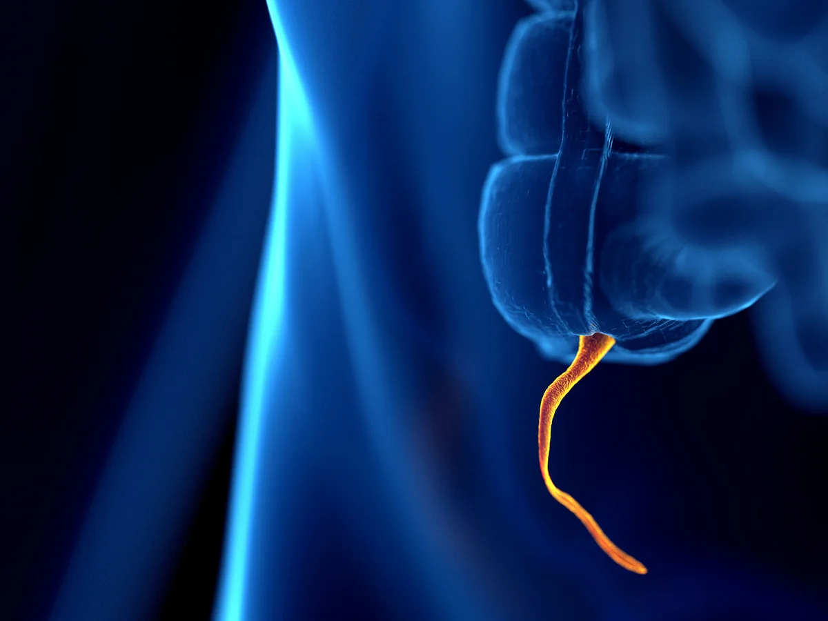
The human appendix is a blind-ended, narrow muscular tube measuring 5 to 15 centimeters in length, arising from the posteromedial border of the cecum. Its luminal orifice is guarded by the valve of Gerlach (a mucosal fold). Colonic peristalsis naturally propels digestive contents upwards into the ascending colon, steering dense debris away from the appendiceal opening.
However, when gravitational forces, body posture, and a wide anatomical orifice align, a heavy swallowed fragment can slip into the appendix. Once inside, retrograde propulsion is almost non-existent due to weak appendiceal smooth muscle peristalsis. Medical epidemiology indicates that foreign bodies cause appendicitis in only 0.005% of all documented surgical cases.
The valve of Gerlach permits heavy food debris to slip inside during low cecal motility, after which retrograde escape is prevented by muscular spasm.
The intestinal mucosa seals the foreign object in dense layers of mucus, indigestible fibers, and calcium salts, shielding tissue from acute infection.
Colicky pain in the absence of fever and elevated inflammatory markers results from physical friction against intraluminal nerve plexuses during peristalsis.
Minimally invasive excision via small keyhole incisions enables safe organ extraction without spilling infected intraluminal contents into the peritoneum.
Comparative Risks: Sharp Objects Versus Calcified Fecalomas
The clinical risk profile of an appendiceal foreign body is largely determined by its sharp contours and physical composition. Pointed needles, toothpicks, or fish bones carry an immediate risk of transmural perforation, often provoking acute localized peritonitis within hours of entry. In contrast, blunt or rounded objects can linger for decades, gradually encased in concentric layers of organic debris and calcium salts.
Medical literature documents a famous historical case where an elderly hunter carried 27 lead shot pellets inside his appendix for over 30 years after eating game fowl, without developing acute inflammation. However, a bone remaining in the appendix for 15 years without histological appendicitis is considered an unprecedented record. For deeper insight into cellular and physiological mechanisms, explore our coverage on the Alzheimer paradox and tau protein dynamics.
- Sharp bones (fish, poultry) warrant urgent medical evaluation due to perforation risks in the upper GI tract within 6 to 12 hours.
- When evaluating recurrent right lower abdominal pain, patients should disclose distant histories of swallowing hard foreign objects.
- Normal blood tests and absence of fever do not rule out a large mechanical fecaloma obstructing the appendiceal lumen.
- High-resolution contrast-enhanced abdominal CT remains the most reliable modality for detecting radio-opaque foreign bodies in the appendix.
Diagnostic Takeaways: The Limitations of Ultrasound and the Vital Role of CT
This extraordinary clinical case underscores the diagnostic limitations of ultrasound in lower quadrant abdominal pain. Intestinal gas in the cecum frequently generates acoustic shadows that obscure the tip of the appendix, obscuring large calcified fecalomas. High-resolution contrast-enhanced CT remains the definitive diagnostic gold standard for evaluating unexplained recurrent abdominal discomfort.
The surgical team highlighted that the patient’s recurrent spasms were triggered by physical friction against intraluminal nerve plexuses during intestinal peristalsis rather than bacterial infection. Timely laparoscopic appendectomy successfully averted eventual wall necrosis, bowel perforation, and secondary sepsis.