



For decades in clinical neurology, phosphorylated tau protein—specifically the p-tau217 isoform—has been regarded as an unequivocal biochemical hallmark of lethal neurodegeneration: a toxic culprit that accumulates inside aging neurons, condenses into insoluble neurofibrillary tangles, and decimates memory networks in Alzheimer’s disease. However, a revolutionary study conducted by Swedish neuroscientists at the University of Gothenburg has overturned this foundational paradigm. Highly sensitive blood plasma assays revealed that the highest concentrations of p-tau217 ever recorded across the human lifespan occur not in end-stage dementia patients, but in perfectly healthy full-term and premature newborn infants. Published in Brain Communications, this unexpected breakthrough proves that this supposed «biomarker of decline» is actually an indispensable driver of early brain development, charting an entirely new therapeutic frontier against dementia.
The Gothenburg Breakthrough in Brain Communications: Lifespan Blood Screening
The research team at the Sahlgrenska Academy of the University of Gothenburg, led by Kaj Blennow and Thomas Karlsson, published findings that caught clinical neurologists completely off guard. Utilizing ultra-sensitive Single Molecule Array (SIMOA) immunoassay platforms, the scientists quantified circulating p-tau217 concentrations across a vast cross-section of human cohorts, spanning premature infants, full-term newborns, young adults, cognitively intact elderly individuals, and diagnosed Alzheimer’s patients.

The data revealed an extraordinary biological phenomenon: blood concentrations of p-tau217 in neonates during their first days of life were multiple times higher than the levels measured in elderly patients suffering from severe Alzheimer’s disease. The highest concentrations of all occurred in premature infants born before 32 weeks of gestation.
Longitudinal tracking over the ensuing months showed that circulating p-tau217 declined steeply, plateauing near standard adult baselines by the end of the first year of life. This provided undeniable proof that tau hyperphosphorylation is a strictly orchestrated, non-pathological physiological feature of early neurodevelopment.
The Biochemistry of Synaptogenesis: Why Growing Axons Demand Cytoskeletal Fluidity
To comprehend why an infant’s brain is flooded with p-tau217, one must inspect the nanomechanics of neuron growth. During late gestation and early infancy, the human brain undergoes an explosive phase of synaptogenesis, generating millions of novel synaptic connections every second. Growing axons and dendrites must elongate, steer toward biochemical cues, and construct intricate neural circuitry spanning both hemispheres.
The structural framework of axons consists of microtubules assembled from tubulin subunits. In mature neurons, dephosphorylated tau binds tightly to microtubules, acting like rigid railroad ties that stabilize the cell’s architecture. However, an unyielding cytoskeleton cannot rapidly elongate or negotiate directional turns. When tau is phosphorylated at the threonine-217 residue, its binding affinity plummets, temporarily freeing the microtubules to disassemble, flex, and reassemble with immense structural plasticity.
Just as profound altered states of consciousness, from the neurobiology of lucid dreaming and deep meditative states to near-death phenomena, recruit extraordinary synaptic flexibility, the nascent brain leverages hyperphosphorylation as an evolutionary shortcut for structural agility.
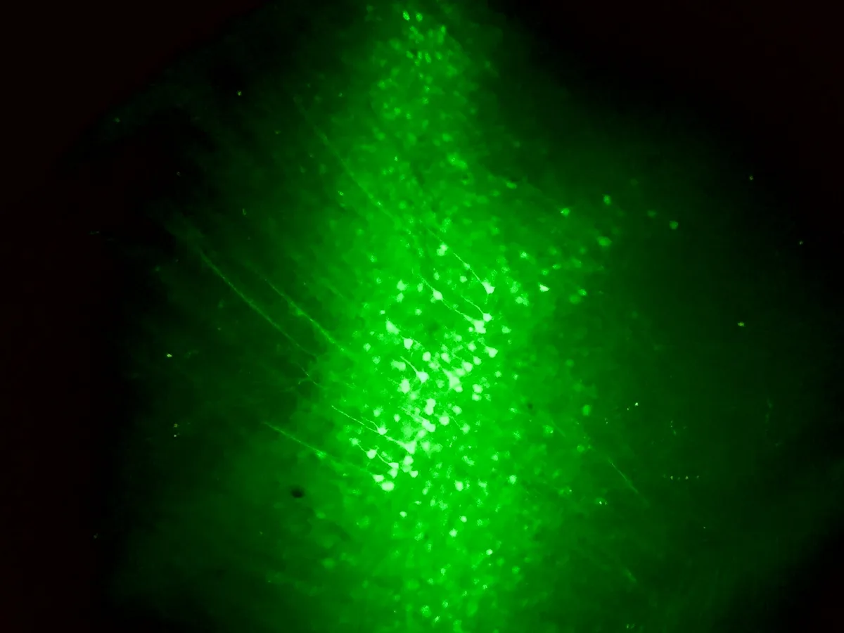
«Discovering stratospheric levels of p-tau217 in newborn babies sent shockwaves through the neuroscience community. We spent years treating this molecule strictly as toxic waste destroying synapses. Now it is undeniable that in the developing infant brain, p-tau217 performs an essential physiological function: enabling the rapid remodeling of the neuronal cytoskeleton as billions of axons wire up. The million-dollar question has fundamentally shifted: how do baby brains safely tolerate massive amounts of this protein without harm, and why does the aging brain lose this shield?»
Kaj Blennow, Professor of Clinical Neurochemistry at the University of Gothenburg and Senior Study Author
Antagonistic Pleiotropy in Action: The Evolutionary Cost of Human Longevity
The Gothenburg findings represent a quintessential real-world demonstration of antagonistic pleiotropy—an evolutionary concept formulated by evolutionary biologist George Williams in 1957. The hypothesis posits that biological mechanisms conferring immense survival advantages during early development and reproductive fitness will be favored by natural selection, even if they inflict severe deleterious consequences during post-reproductive senescence.
For the developing embryo, the high structural plasticity driven by p-tau217 is essential for survival, enabling the rapid expansion of the neocortex. However, when the very same biochemical phosphorylation pathways are reactivated late in life—triggered by neuroinflammation, metabolic dysfunction, or beta-amyloid accumulation—the outcome is disastrous.
In an aging neuron, detached phosphorylated tau molecules find no growing axons to support. Instead, they aggregate into soluble toxic oligomers, coalesce into paired helical filaments, and ultimately deposit as insoluble neurofibrillary tangles that choke cellular transport machinery, leading to apoptosis and severe cognitive collapse.
Circulating p-tau217 levels in preterm infants (<32 weeks) exceed the concentrations of late-stage Alzheimer's patients by 2 to 3 times.
Biomarker levels decline exponentially throughout the first year of life as the initial wave of axonal sprouting reaches completion.
Phosphorylation at threonine-217 temporarily releases tau from tubulin, enabling rapid branching and growth cone steering.
An adaptive pathway that builds the infant brain early in life becomes an engine of toxic aggregation during late-life senescence.
The Newborn Shield: How Infant Brains Evade Neurofibrillary Toxicity
The pivotal challenge facing neuropharmacologists today is clear: why do infant brains, despite harboring three to four times more p-tau217 than Alzheimer’s patients, never develop neurofibrillary tangles or neurodegeneration? Investigators have isolated three primary endogenous protective systems operating in the infant central nervous system.
First, neonatal neurons exhibit hyperactive molecular chaperone networks, specifically heat shock proteins HSP70 and HSP90, alongside robust ubiquitin-proteasome systems that isolate monomeric phospho-tau, preventing liquid-liquid phase separation (LLPS) from crystallizing into amyloid seeds. Second, neonatal microglia and astrocytes reside in a pro-reparative phenotype, continuously cleansing the extracellular matrix without producing destructive pro-inflammatory cytokines.
Studying the boundary conditions of neural tissue consistently yields profound clinical breakthroughs—much like exploring how shared death experiences shed light on terminal neural synchronization at the edge of life, understanding the newborn brain’s natural immunity to tau pathology provides the master blueprint for arresting Alzheimer’s disease.
- Phosphorylated tau p-tau217 is not an exclusive biomarker of dementia; it is a vital physiological driver of early synaptogenesis.
- Infant brains produce potent molecular chaperones and proteasomes that completely block tau from condensing into lethal tangles.
- Future Alzheimer’s therapies should focus on emulating infant chaperone resilience rather than relying solely on protein eradication.
- Neonatal p-tau217 tracking represents a revolutionary objective biomarker for pediatric brain maturation and neurological health.
Therapeutic Horizons: Moving Beyond Protein Clearance to Emulating Infant Resilience
Current pharmaceutical approaches (such as lecanemab and donanemab) have concentrated nearly exclusively on monoclonal antibodies engineered to clear extracellular amyloid or tau plaques. However, these therapies exhibit modest clinical efficacy and carry dangerous risks of amyloid-related imaging abnormalities (ARIA), including cerebral microhemorrhages and localized vasogenic edema.
The Swedish breakthrough offers an inspiring alternative: instead of indiscriminately purging tau protein from the brain, therapies should aim to pharmacologically reactivate the chaperone and lysosomal degradation networks that maintain tau solubility in neonates. If aging neurons can be coaxed into preserving phosphorylated tau in its monomeric, fluid state, tangle formation would halt permanently, even in the presence of elevated biomarker levels.
Moreover, pediatricians have gained a transformative diagnostic instrument: tracking the rate of p-tau217 clearance during the first year of life offers an objective biomarker to detect perinatal hypoxia, white matter injury, and neurodevelopmental delays years before behavioral manifestations emerge.