

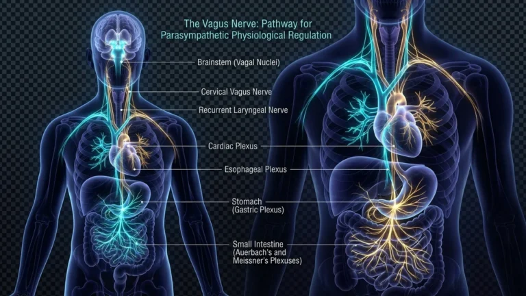

Spanning the core of human physiology lies the longest and most pervasive cranial pathway of the parasympathetic nervous system: the vagus nerve (Cranial Nerve X). Originating within the brainstem, it descends through the cervical sheath and thorax to innervate the heart, lungs, liver, and gastrointestinal tract. The discovery of the cholinergic anti-inflammatory pathway transformed vagal tone from an abstract physiological concept into the vanguard of evidence-based neuroimmunology, establishing the vagus nerve as our primary internal brake against systemic panic and chronic inflammatory decay.
Anatomy of Cranial Nerve X: From the Medulla to Enteric Plexuses
Earning its Latin moniker vagus (“wandering”) due to its expansive visceral distribution, the nerve emerges from the medullary olivary nucleus and traverses the jugular foramen. Descending within the carotid sheath, it branches extensively into recurrent laryngeal, pulmonary, cardiac, celiac, and mesenteric plexuses, terminating near the splenic flexure of the colon.
Decades of anatomical re-evaluation overturned the assumption that the vagus functions solely as an executive motor brake. Approximately 80% of its axon bundle consists of unmyelinated sensory afferents. These fibers continuously monitor aortic baroreceptor stretch, gastric distention, and mucosal cytokine concentrations, feeding real-time somatic metrics into the nucleus tractus solitarius (NTS).
The Cholinergic Anti-Inflammatory Pathway: Kevin Tracey’s Breakthrough
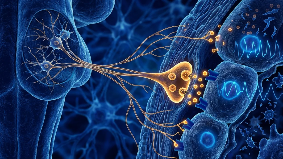
In landmark investigations spearheaded by neurosurgeon Dr. Kevin Tracey, researchers established that the central nervous system exerts direct molecular control over peripheral immune cascades. When splenic macrophages overproduce tumor necrosis factor (TNF-alpha) during sepsis or chronic inflammatory states, vagal stimulation initiates splenic nerve activation and acetylcholine release.
Acetylcholine binds selectively to alpha-7 nicotinic acetylcholine receptors (alpha-7 nAChR) on macrophage membranes, suppressing transcription factor NF-kB translocation and halting pro-inflammatory cytokine transcription. High vagal tone operates as a vital endogenous prophylactic against atherosclerosis, metabolic syndrome, and cellular senescence.
Sensory fibers transmit visceral organ metrics upward to the medullary NTS.
Acetylcholine binding to alpha-7 nAChR suppresses macrophage cytokine synthesis.
Elevated interbeat variability mirrors parasympathetic resilience and stress recovery.
Breathing with extended exhalations engages mechanical baroreceptor braking.
Heart Rate Variability (HRV): The Gold Standard Biomarker of Vagal Tone
In clinical cardiology and sports science, parasympathetic regulatory efficiency is quantified via Heart Rate Variability (HRV)—the millisecond fluctuations separating consecutive R-R interbeat intervals. A resilient heart does not fire like a metronome; during inhalation, sympathetic acceleration predominates, while exhalation elicits instantaneous vagal acetylcholine release, slowing sinus nodal discharge (Respiratory Sinus Arrhythmia).
Elevated resting root mean square of successive differences (RMSSD) reflects robust vagal brake capacity. Cohorts exhibiting high baseline HRV demonstrate superior neuroendocrine recovery from acute psychosocial stress, lower systemic C-reactive protein, and enhanced executive cognitive control.
«The vagus nerve is an expansive bi-directional communication superhighway where up to 80% of fibers are afferent—transmitting sensory surveillance data from visceral organs upward to the brainstem. Emotional resilience is not forged through sheer mental willpower, but through somatic regulatory loops mediated by vagal circuitry.»
Dr. Stephen Porges, Distinguished University Scientist and author of The Polyvagal Theory
Evidence-Based Somatic Protocols: Diaphragmatic Breath and Cold Shock
Because vagal branches innervate the diaphragm, vocal cords, and facial sensory arches, individuals can directly modulate vagal outflow through targeted non-pharmacological interventions. Slow diaphragmatic breathing featuring prolonged exhalations (4 seconds in, 7–8 seconds out) expands alveolar mechanoreceptors, triggering baroreflex-mediated parasympathetic discharge.
Simultaneously, the mammalian dive reflex—elicited by immersing the face in cool water (10–12°C)—activates trigeminal-vagal pathways, inducing rapid bradycardia and peripheral vasoconstriction. Vocal exercises including singing, rhythmic humming, and deep gargling mechanically stimulate the recurrent laryngeal nerve, fortifying resting vagal tone.
- Deploy the physiological sigh: two quick nasal inhalations followed by a slow, complete oral exhalation.
- Incorporate brief facial immersion in cold water to trigger the parasympathetic diving reflex.
- Engage in consistent Zone 2 aerobic training to heighten autonomic baroreflex responsiveness.
- Utilize vocal vibration practices such as deep humming and gargling to stimulate laryngeal vagal branches.
Bioelectronic Horizons: VNS Implants and Non-Invasive Neuromodulation
Vagus Nerve Stimulation (VNS) has advanced into an FDA-approved clinical therapy for refractory major depression and intractable epilepsy. Surgically implanted pulse generators deliver low-frequency electrical currents along the left cervical vagus trunk, stimulating locus coeruleus noradrenaline release and dorsal raphe serotonergic transmission.
Concurrently, transcutaneous auricular vagus nerve stimulators (taVNS) targeting the cymba conchae (Arnold’s nerve) demonstrate remarkable clinical efficacy in alleviating post-traumatic stress disorder, generalized anxiety, and chronic migraine syndromes without invasive surgical risks.