



For hundreds of years, medical textbooks and anatomical engravings contained a peculiar and telling blind spot: the pregnant human body was almost universally depicted without a head — merely a headless torso featuring a uterus, placenta, and growing fetus, as if everything above the neck remained entirely untouched by gestation. Yet contemporary neuroscience has established that how motherhood transforms the brain represents the most profound, rapid, and enduring wave of neuroplasticity experienced by the adult human nervous system. Landmark longitudinal studies led by neuroscientist Dr. Susana Carmona of the Gregorio Marañón Health Research Institute in Madrid prove that pregnancy triggers a sweeping architectural reorganization that surpasses even adolescent development, recalibrating a woman’s cognitive networks, emotional circuits, and social cognition for decades to come.
Centuries of Anatomical Neglect: Why Medicine Left the Pregnant Patient Headless
Examine the historical trajectory of medical illustrations from the Renaissance through the late twentieth century, and a striking pattern emerges: pregnant women were routinely rendered as headless torsos. Medical curricula meticulously mapped the physiological adjustments of nearly every visceral organ: cardiac output surging by 40–50%, plasma volume expanding, glomerular filtration accelerating, and the maternal immune system undergoing sophisticated modulation to tolerate semi-allogeneic fetal tissues.

This profound physiological understanding enabled clinicians to identify and manage life-threatening pathologies such as preeclampsia and gestational diabetes. Yet the brain — the central regulator of neuroendocrine homeostasis and human behavior — was conspicuous by its absence. Generations of clinicians were trained to perceive pregnancy as an exclusively pelvic process, leaving maternal cognitive and mental health relegated to folk myths.
Previously, we explored how functional neuroimaging unlocks cortical processing in our analysis of neuroimaging and fMRI decoding of neural networks. Today, these exact neuroimaging technologies are illuminating maternal brain reorganization.
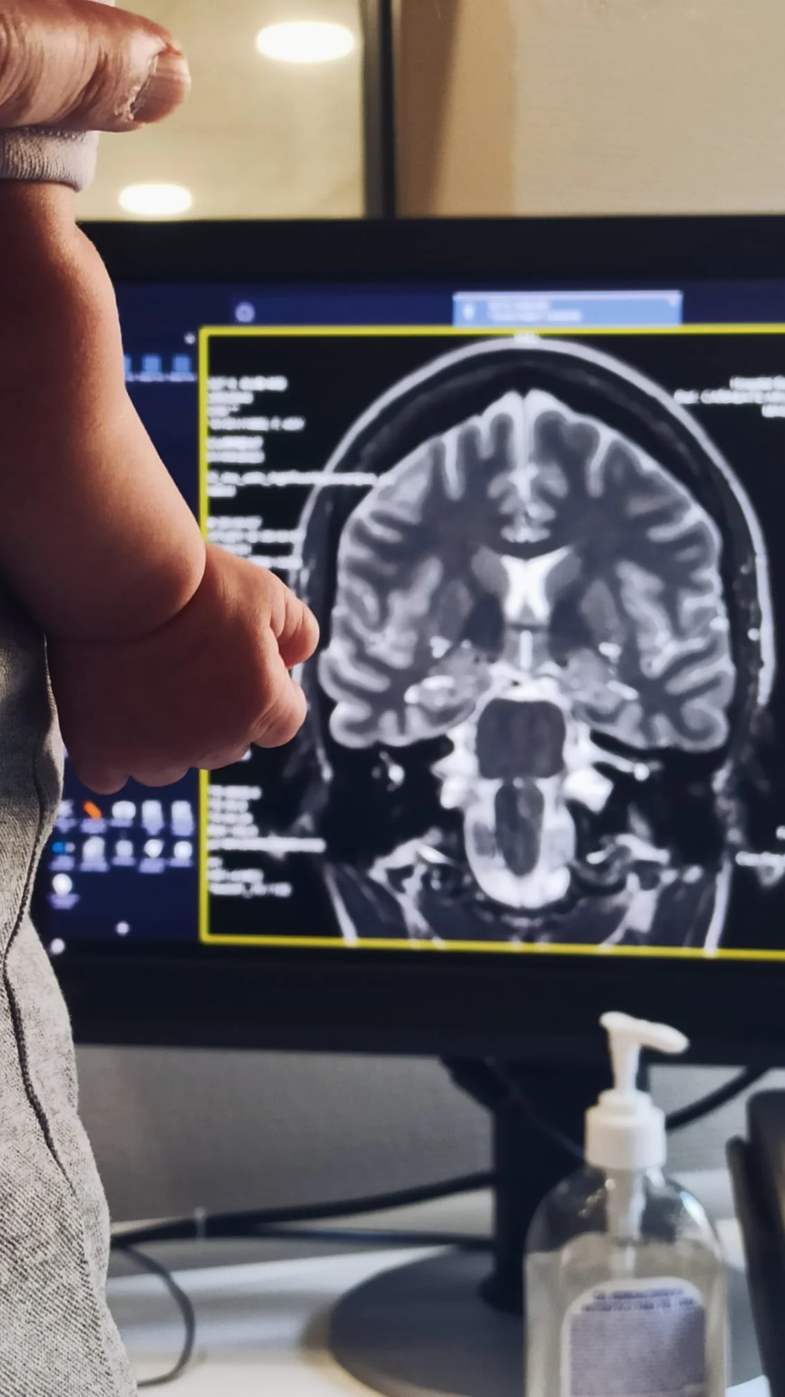
It was not until 2016 that an international team led by Dr. Susana Carmona and Dr. Elseline Hoekzema published the first prospective MRI study in Nature Neuroscience demonstrating that pregnancy profoundly and systematically reorganizes the anatomical architecture of the human cerebral cortex.
Neuroplasticity on an Unprecedented Scale: Synaptic Pruning Rather Than Brain Shrinkage
The central finding of pre- and post-pregnancy magnetic resonance morphometry is a widespread, symmetrical reduction in gray matter volume across the cerebral cortex. Initial media coverage sensationalized these findings, mistakenly declaring that pregnancy «shrinks» or degrades maternal cognitive faculties.
Modern cellular neuroscience clarifies that this volume reduction represents a textbook manifestation of synaptic pruning. In a mechanism identical to the pubertal remodeling of the adolescent brain, microglia and astrocytes eliminate redundant, non-functional synaptic spines while strengthening core transmission pathways, rendering neural circuits substantially more specialized and energetically efficient.
As Dr. Carmona articulates in her groundbreaking book «A Mother’s Brain: The New Science of the Neuro-Maternal Revolution» (The MIT Press), this architectural recalibration is akin to upgrading a computer’s processor: streamlining connections to dramatically accelerate complex real-time computational performance.
The structural changes are predominantly localized within the Default Mode Network (DMN) and social cognition networks responsible for Theory of Mind — the mental capacity to infer another individual’s emotional states, intentions, and unspoken distress signals, a survival prerequisite when caring for a non-verbal infant.
«That blank space above the neck in medical textbooks was not an oversight by the illustrator. It is a faithful map of what medicine chose to study. We now have the tools to fill in what we left blank.»
Dr. Susana Carmona, Director of the Neuromaternal Lab at the Gregorio Marañón Health Research Institute, author of «A Mother’s Brain» (The MIT Press)
Debunking the Myth of «Mommy Brain»: Evolutionary Adaptation vs. Exhaustion
A pervasive cultural trope claims that pregnancy induces «momnesia» or «mommy brain» — a colloquial term used to describe transient absentmindedness, misplaced objects, and subjective brain fog. For decades, this phenomenon was casually dismissed as an inevitable cognitive impairment.
However, rigorous neuropsychological testing paints a completely different picture. In controlled cognitive batteries evaluating general intelligence, semantic retrieval, and problem-solving, mothers do not exhibit significant intellectual deficits. On the contrary, post-partum women demonstrate superior vigilance, faster threat detection, and heightened accuracy in decoding micro-facial emotional cues.
Subjective reports of absentmindedness are the product of profound environmental and physiological factors: chronic sleep fragmentation, the massive metabolic costs of lactation, and intense selective attentional tuning. The maternal brain deliberately filters out irrelevant peripheral minutiae to channel 100% of cognitive bandwidth into infant survival.
Crucially, longitudinal neuroimaging has shown that the magnitude of cortical gray matter reduction directly correlates with maternal-infant bonding metrics: mothers exhibiting the most pronounced structural remodeling report warmer, more intuitive emotional attachment with their newborns.
The reduction in cortical gray matter volume reflects cellular fine-tuning rather than cognitive decline, eliminating extraneous synapses to optimize maternal responsiveness.
Cortical reorganization selectively concentrates in DMN hubs, transforming self-referential mentalization into heightened empathic attunement toward the infant.
Standardized neuropsychological testing disproves cognitive deficits: perceived brain fog stems from sleep fragmentation, while threat vigilance and memory recall are sharpened.
Heightened neural plasticity renders the brain uniquely vulnerable: perinatal depression affects 1 in 5 women, making postpartum suicide a leading cause of maternal mortality.
The Dark Side of Plasticity: The Window of Biological Vulnerability and Perinatal Depression
High neuroplasticity is an evolutionary double-edged sword. A nervous system undergoing rapid structural flux is extraordinarily adaptable, but it is also far more susceptible to systemic disturbances, chronic distress, social isolation, and physiological dysregulation.
In a landmark 2025 study from the Neuromaternal Laboratory published in Science Advances, researchers documented that atypical trajectories in postpartum cortical recovery correlate with the onset and severity of peripartum affective disorders.
Epidemiological data underscore the gravity of this vulnerability: roughly 1 in 5 women experience clinical perinatal depression or anxiety. In developed nations, suicide linked to peripartum mental illness ranks as one of the leading causes of maternal mortality within the first year after childbirth.
When societal narratives dismiss this period as merely joyful while failing to provide institutional, clinical, and familial support, the consequences can be catastrophic. The maternal brain requires rigorous preventative monitoring, destigmatized psychiatric care, and comprehensive community backing.
- Pregnancy drives the most extensive structural brain reorganization of adulthood, outstripping adolescent neuroplasticity in both speed and intensity.
- The degree of cortical gray matter pruning directly predicts the strength and quality of mother-infant emotional bonding.
- Perinatal mental health disorders represent an urgent global public health priority, demanding dedicated open multi-modal longitudinal datasets.
A Call for a Global Neuro-Maternal Revolution: Open Data Biobanks and AI Diagnostics
The foremost obstacle impeding progress in maternal healthcare is data fragmentation. Fetal biobanks, maternal hormone panels, postpartum neuroimaging scans, and social demographic records currently exist in isolated academic silos across different institutions, rarely aggregated into unified longitudinal pipelines.
Major neurodegenerative disorders possess multi-center data consortia like the Alzheimer’s Disease Neuroimaging Initiative (ADNI), while adolescent cognitive development is tracked through the Adolescent Brain Cognitive Development (ABCD) study. Motherhood — an experience shared by 85% of women that underpins the perpetuation of our species — urgently warrants an equivalent open, longitudinal research infrastructure.
As evidenced by recent breakthroughs where functional neuroimaging and cognitive cortex mapping empower machine learning models, integrating multi-modal biomarkers will enable artificial intelligence to forecast postpartum depression risks during early pregnancy.
It is time to permanently retire the headless depictions of the pregnant patient. Giving the maternal brain its rightful place in scientific inquiry is essential for maternal survival and the thriving of future generations.