


A diagnostic rarity documented fewer than four hundred times across four centuries of global medical history has astonished surgeons in the Horn of Africa. Published in the peer-reviewed International Medical Case Reports Journal, an extraordinary clinical report details the case of a 42-year-old woman in Somaliland who endured three years of persistent, dull lower abdominal pain and unexplained pelvic fullness. Advanced three-dimensional computed tomography (3D-CT) unraveled the startling etiology: an 18-by-13-centimeter calcified extrauterine mass resting within her peritoneal cavity—a classic lithopedion, colloquially known as a ‘stone baby,’ originating from an undetected primary abdominal ectopic pregnancy.
Chronic Abdominal Discomfort: Patient Presentation and Clinical History
In early 2026, a 42-year-old multiparous woman (gravida 4, para 3) presented to the emergency surgical service at Hargeisa Group Hospital in Somaliland, reporting a three-year history of worsening, dull lower abdominal pain and an unrelenting sensation of pelvic fullness. Her symptoms were accompanied by intermittent obstipation, progressive dyspepsia, and urinary urgency, severely diminishing her daily quality of life.
A meticulous clinical history revealed a crucial obstetric clue: three years earlier, the patient had believed herself to be pregnant. She experienced early morning nausea, progressive abdominal enlargement, and unequivocal quickening (fetal movements) during the second trimester. Several months later, however, fetal movements abruptly ceased, followed by a slight decrease in abdominal girth without labor contractions or vaginal hemorrhage. Residing in a remote rural locale with limited obstetric sonography infrastructure, she sought no medical evaluation, assuming the pregnancy had spontaneously dissolved.
Upon physical palpation, the surgical team detected a firm, non-tender, stone-hard mass occupying the suprapubic region and right iliac fossa. It measured roughly the dimensions of an infant’s head and was moderately mobile. Initial differential diagnoses included a heavily calcified subserosal uterine leiomyoma (fibroid), a mature benign ovarian teratoma, or an ancient hydatid echinococcal cyst.
Cross-Sectional Imaging: 3D-CT Reveals the Skeletal Remains
Initial transabdominal ultrasonography produced confounding findings: the uterus was completely empty with an unremarkable endometrial stripe, and bilateral adnexa demonstrated no focal cystic or solid pathology. Directly cephalad to the uterine fundus, however, sonographers encountered a dense curvilinear acoustic shadow completely attenuating sound wave transmission.
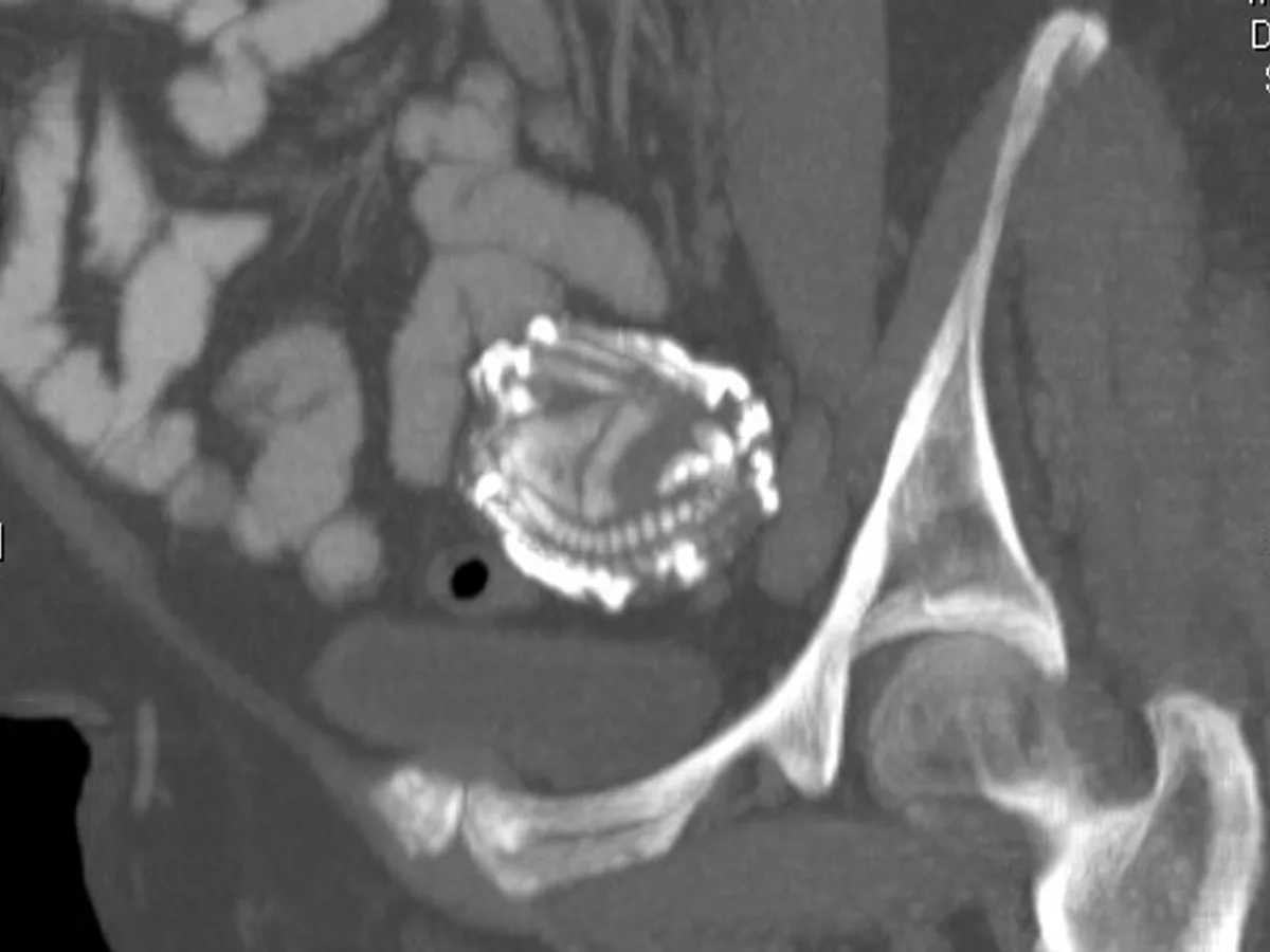
To achieve diagnostic certitude, the medical team ordered contrast-enhanced multidetector computed tomography (MDCT) of the abdomen and pelvis paired with volume-rendered 3D reconstructions. The radiologic findings were definitive and startling: nestled within the peritoneal cavity between the anterior abdominal wall, the uterine fundus, and the ileal mesentery lay a well-circumscribed, radio-dense mass measuring 18.2 × 13.1 × 11.4 cm.
Within the hyper-dense, calcified outer shell, CT clearly depicted the unmistakable architectural elements of a late-term fetal skeleton. The calvarium exhibited patent cranial sutures, while the thoracic cage, segmented vertebral column, pelvis, and elongated femur and humerus bones were preserved in a classic flexed intrauterine posture. The diagnosis was unambiguous: an abdominal lithopedion.
Pelvic cross-sectional imaging revealed a fully developed fetal skeleton complete with cranium, ribs, and extremities preserved inside an eggshell-like calcium matrix.
Lithopedion is among the rarest conditions known to obstetric surgery, with the first scientifically validated postmortem examination recorded in France in 1582.
The fertilized blastocyst implanted directly onto the peritoneal mesentery, drawing blood supply from visceral vessels rather than the uterine endometrium.
Surgical dissection successfully mobilized the densely adherent mass away from the small bowel and urinary bladder, completely resolving the patient’s chronic pain.
Pathogenesis: How the Immune System Mummifies Extrauterine Life
Derived etymologically from the Greek terms lithos (stone) and paidion (child), a lithopedion represents an extraordinary biological adaptation to an otherwise catastrophic reproductive event. In normal biology, non-viable conceptuses within the Fallopian tube or uterus undergo enzymatic autolysis, leukocyte infiltration, and spontaneous resorption or expulsion.
In approximately 1 in 11,000 gestations, however, an advanced extrauterine abdominal pregnancy occurs. Following primary peritoneal implantation or early secondary extrusion after tubal rupture, the trophoblast adheres to the parietal peritoneum, omentum, or mesenteric arcade, maintaining vascularization sufficient to sustain fetal viability well into the second or third trimester.
When the fetus eventually succumbs to placental insufficiency or hypoxia beyond 14 weeks of gestation, its physical volume overwhelms maternal macrophage clearance mechanisms. To forestall fatal liquefactive necrosis, polymicrobial peritonitis, and maternal sepsis, the host immune response initiates a foreign-body encapsulation reaction. Dense fibrous connective tissue seals off the dead fetus, followed by dystrophic calcification—the relentless precipitation of calcium phosphate and carbonate salts. Over months and years, the organic tissue is petrified into a sterile, mineralized relic.
«A lithopedion represents an astonishing biological defense mechanism. When a late-gestation fetus perishes outside the womb, the maternal immune system recognizes it as a foreign body and encases it in calcium salts, shielding the patient from lethal septic shock.»
Dr. Abdisamed Mohamoud H Ali, Lead Surgeon, Hargeisa Group Hospital & Amoud University
Historical Echoes: From Albucasis to Madame Colombe Chatri
The historical medical literature on lithopedia is remarkably scarce, reflecting the sheer rarity of surviving advanced untreated abdominal pregnancies. The earliest recorded historical allusion appears in the works of the 10th-century Moorish surgeon Abu al-Qasim al-Zahrawi (Albucasis) of Córdoba, who documented the postmortem extraction of a petrified fetal mass from an abdominal incision.
The first systematically validated scientific autopsy was published in 1582 in Sens, France. Renowned physician Jean d’Aassigny performed a postmortem examination on 68-year-old Madame Colombe Chatri, discovering a perfectly preserved, calcified fetal mass that had resided in her pelvic cavity for 28 years following an obstructed labor in 1554. Dubbed the ‘Lithopedion of Sens,’ the specimen caused a scientific sensation and was purchased by King Christian III of Denmark.
Over the past four centuries, fewer than 400 authenticated cases have been documented in peer-reviewed journals globally. While modern antenatal ultrasound screening has made the condition vanishingly rare in developed nations, it occasionally surfaces in medically underserved populations, with documented retention intervals extending up to six decades.
- Prerequisite threshold: fetal development must progress past 14 weeks of gestation, exceeding the phagocytic clearance capacity of maternal macrophages;
- Immune walling-off: triggering of intense fibroblastic encapsulation followed by dystrophic calcification (calcium phosphate and hydroxyapatite deposition);
- Clinical classification: lithokelyphos (calcification restricted to gestational membranes), true lithopedion (fetus itself calcified), and lithokelyphopedion (both calcified);
- Diagnostic pitfalls: on pelvic ultrasound, the acoustic shadowing mimics a calcified uterine leiomyoma, benign cystic teratoma, or hydatid cyst.
Surgical Laparotomy and Patient Recovery: Clinical Lessons
Although lithopedia can remain clinically quiescent for decades, they carry formidable latent morbidity, including bowel fistulization, recurrent pelvic abscesses, and mechanical intestinal obstruction. Given the Somaliland patient’s persistent, debilitating pain, operative excision was unequivocally indicated.
A surgical team led by Dr. Abdisamed Mohamoud H Ali performed an exploratory lower midline laparotomy. Intraoperatively, the calcified mass was found densely bound by vascularized fibrous adhesions to the serosa of the terminal ileum and the peritoneal reflection of the urinary bladder. Through meticulous sharp and blunt dissection, the surgeons preserved bowel wall integrity, successfully extirpating the stone mass in its entirety.
The patient experienced an uneventful postoperative recovery, regaining normal gastrointestinal function by postoperative day two and achieving complete, lasting relief from the pelvic agony that had compromised her life for three years. This compelling clinical outcome underscores the importance of maintaining a broad diagnostic index of suspicion when evaluating complex pelvic calcifications, serving as a powerful testament to the defensive adaptations of the human body.