

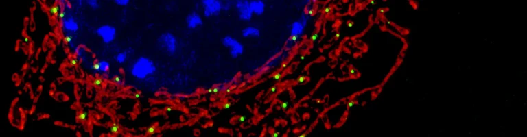

For over a century, classical cell biology viewed eukaryotic cells as discrete, autonomous entities that communicate solely through soluble paracrine signals or extracellular vesicles. However, the discovery of Tunneling Nanotubes (TNTs) revolutionized contemporary cytology: cells are capable of projecting direct, continuous membranous conduits spanning tens to hundreds of micrometers. Along these intercellular highways, donor cells — most notably mesenchymal stem cells (MSCs) — execute horizontal transfer of intact organelles, preferentially healthy and bioenergetically active mitochondria. Upon entering hypoxic neurons or ischemic cardiomyocytes, these donor mitochondria immediately fuse with host networks, restore ATP synthesis, and miraculously rescue dying tissues.
Architecture of Tunneling Nanotubes: Direct Cytoplasmic Superhighways
The phenomenon of tunneling nanotubes was first uncovered in 2004 by the pioneering research team led by Professor Hans-Hermann Gerdes. They documented ultra-thin, continuous membranous bridges ranging from 50 to 800 nanometers in diameter, extending across vast intercellular spaces.
Unlike traditional gap junctions, which permit only the exchange of small metabolites under 1.5 kilodaltons, TNTs are open cylindrical corridors enveloped by a continuous phospholipid bilayer that directly fuses the cytoplasmic contents of two distant cells.
The internal structural scaffolding of these conduits is forged by polymerised filamentous F-actin, stabilized by unconventional myosins (Myo10 and Myo19). This actin core confers tensile strength and provides directional molecular rails for transport.
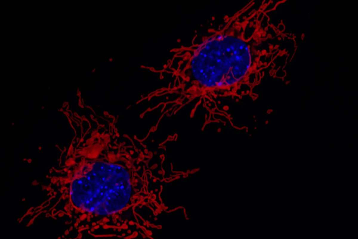
The Machinery of Organelle Transit: The Miro1 GTPase and Milton Adaptor Complex
Trafficking an organelle as massive as a mitochondrion through a submicron channel demands sophisticated molecular mechanics. The principal motor coordinator governing this transit is Miro1 (Rhot1), an atypical Rho-GTPase embedded in the outer mitochondrial membrane.
Miro1 binds to Milton adaptor proteins (TRAK1 and TRAK2), forming a stable tripartite complex that anchors to the heavy chains of Kinesin-1 (KIF5B). Driven by ATP hydrolysis, kinesin motors step along internal cytoskeletal tracks, ferrying the mitochondrion at speeds of 1 to 2 micrometers per minute.
Experimental upregulation of Miro1 dramatically augments mitochondrial donation rates, whereas genetic knockout of Miro1 cripples organelle migration, cementing Miro1 as the indispensable molecular master switch of metabolic rescue.
In addition to Miro1, the outer mitochondrial membrane dynamin-related GTPase Mitofusin-2 (Mfn2) coordinates organelle remodeling prior to translocation. By transiently promoting mitochondrial fission, cells downsize organelles to fit through the narrow actin lumen, enabling smooth passage before re-fusing into active tubular networks upon recipient entry.
Direct membranous cylinders (50–800 nm diameter) bridging the cytoplasm of separate cells into an open syncytium.
Outer membrane GTPase Miro1 couples mitochondria to kinesin-1 motors for active transit along F-actin tracks.
Imported functional mitochondria rapidly revive ATP production in ischemic cells, arresting apoptotic cascade.
Malignant cells extend TNTs to deplete mitochondria from immune T-lymphocytes, evading antitumor destruction.
Metabolic Rescue: Resuscitating Ischemic Tissues in Stroke and Myocardial Infarction
The fundamental physiological stimulus initiating TNT morphogenesis is acute cellular distress and bioenergetic collapse. When cerebral cortical neurons or cardiac myocytes experience severe ischemia, their inner mitochondrial membrane potential collapses, ATP production plummets, and reactive oxygen species (ROS) surge.
Distressed cells release distress chemokines and display phosphatidylserine residues, signaling adjacent mesenchymal stem cells or pericytes to extend filopodial projections and establish TNT conduits. Down these bridges, donor cells deliver functional mitochondria bearing intact respiratory chain complexes.
Once assimilated into the recipient cell, donor mitochondria fuse with endogenous fragments. This rapid integration restores ATP pools, alleviates oxidative stress, closes mitochondrial permeability transition pores, and aborts apoptotic cell death programs.
«Horizontal mitochondrial transfer via tunneling nanotubes demonstrates that tissues operate as a dynamic metabolic syncytium. Cells do not perish in isolation from energetic starvation — they share their powerhouses with suffering neighbors.»
Hans-Hermann Gerdes, Professor of Cell Biology at the University of Bergen
Therapeutic Horizons: Mitochondrial Transplantation in Regenerative Medicine
The deciphering of horizontal mitochondrial transfer has laid the groundwork for an entirely new therapeutic frontier: mitochondrial transplantation therapy. In animal models of acute myocardial infarction, grafting MSCs engineered to overexpress Miro1 reduced fibrotic scar area and preserved ejection fraction.
In models of ischemic stroke, infusing healthy donor mitochondria rescued penumbral neurons, preserving cognitive and locomotor function. Parallel therapeutic triumphs have been documented in acute respiratory distress syndrome (ARDS) and renal tubular injury.
Leading translational laboratories are currently developing cell-free protocols: purifying viable mitochondria from umbilical cord-derived MSCs and delivering them targetedly into damaged organs using biocompatible lipid nanoparticles or cell-penetrating peptides.
- TNTs form within minutes through dynamic localized polymerization of F-actin driven by Myo10 motor proteins.
- Mesenchymal stem cells act as the premier biological donors of mitochondria throughout stressed mammalian tissues.
- Genetic enhancement of Miro1 significantly multiplies the regenerative and survival capacity of transplanted stem cells.
- Direct mitochondrial transplantation is already entering early-phase clinical evaluations for acute cardiac ischemic injury.
The Dark Side of TNTs: How Cancer Cells Hijack Host Energy Reserves
Despite its extraordinary regenerative promise, the tunneling nanotube axis possesses a sinister pathology. Malignant neoplasms frequently hijack this intercellular bridge to bolster their own survival and resist cytotoxic chemotherapy.
Investigations led by Dr. Srirama Venkatesh at Harvard Medical School revealed that aggressive breast cancer and glioblastoma cells form extensive TNT networks with infiltrating cytotoxic CD8+ T lymphocytes. Instead of altruistic sharing, cancer cells siphon active mitochondria away from immune cells.
This organellar piracy leaves antitumor T cells metabolically crippled and exhausted, while cancer cells gain an energetic boost that accelerates invasion and metastasis. Developing pharmacological TNT inhibitors to sever these conduits within the tumor microenvironment is emerging as a compelling immunotherapeutic strategy. Читайте также наш связанный материал про groundbreaking milestones in molecular biology.