

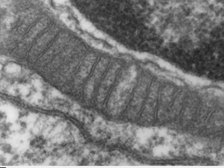

Mitophagy and cellular quality control represent the body’s ultimate molecular defense against aging and neurodegenerative decline: the timely recycling of worn-out mitochondria preserves tissue youth and shields cerebral neurons from cell death. Mitochondria continuously generate cellular adenosine triphosphate (ATP), but during oxidative phosphorylation, they inevitably suffer oxidative damage from free radicals, requiring rigorous intracellular clearance governed by the PINK1 and Parkin signaling pathway.
Mitochondria Under Surveillance: The PINK1-Parkin Kinase Cascade
Every living eukaryotic cell hosts hundreds to thousands of mitochondria arranged within an interconnected dynamic bioenergetic network. Healthy mitochondria maintain a robust negative mitochondrial membrane potential (ΔΨm). However, as oxidative mutations accumulate within mitochondrial DNA (mtDNA) and electron transport complexes falter, the membrane depolarizes, turning the organelle into a hazardous source of reactive oxygen species (ROS).
To avoid triggering apoptotic cytochrome c release and subsequent whole-cell death, selective autophagy—mitophagy—is immediately mobilized. The primary molecular sentinel is PINK1 (PTEN-induced kinase 1). In functional mitochondria, PINK1 is imported across inner membranes and rapidly degraded. Upon depolarization, import halts, stabilizing PINK1 on the outer mitochondrial membrane where it phosphorylates ubiquitin and recruits cytosolic Parkin.
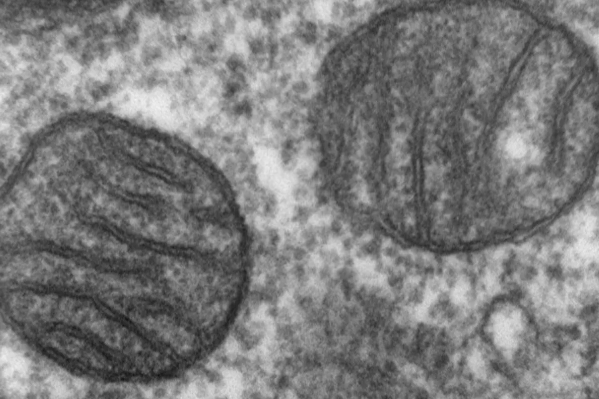
Autophagosome Engulfment and Lysosomal Degradation
Activated Parkin functions as an E3 ubiquitin ligase, extensively decorating damaged outer membrane proteins with polyubiquitin tags. Autophagy adapter proteins (such as optineurin and p62/SQSTM1) recognize these markers, simultaneously binding LC3 proteins located upon the nascent crescent-shaped isolation membrane (phagophore).
The phagophore encapsulates the dysfunctional mitochondrion, sealing it inside a dedicated autophagosome vesicle. This structure then fuses with an acidic lysosome, where specialized hydrolases dismantle the organelle into elemental amino acids, lipids, and nucleotides, allowing the cell to recycle resources into fresh, healthy mitochondria.
Detects membrane depolarization and phosphorylates outer-surface ubiquitin to initiate the clearance cascade.
Coats damaged mitochondrial surfaces with polyubiquitin chains, targeting them for autophagosomal engulfment.
Shields dopaminergic neurons in Parkinson’s pathology and attenuates microglial neuroinflammation in Alzheimer’s disease.
Intermittent fasting, high-intensity aerobic exercise, and Urolithin A powerfully upregulate mitochondrial turnover.
Halting Parkinson’s, Alzheimer’s, and Sarcopenia
Impaired mitophagy is now recognized as a central driver across age-related diseases. Inherited mutations in PINK1 or PRKN cause the accumulation of damaged mitochondria within substantia nigra dopaminergic neurons, precipitating early-onset Parkinson’s disease. In Alzheimer’s pathology, defective mitophagy prevents the removal of dysfunctional mitochondria, exacerbating amyloid-beta plaque accumulation.
«Preserving pristine mitochondrial quality control via mitophagy represents the cornerstone of long-term neuronal resilience and systemic metabolic health.»
Dr. Richard Youle, Breakthrough Prize Laureate in Life Sciences, National Institutes of Health
Within musculoskeletal tissue, waning mitophagy contributes directly to sarcopenia—the progressive decline in skeletal muscle mass and strength observed during aging. Restoring mitophagy reinstates energetic efficiency across muscle fibers. Dive deeper into fundamental life sciences in our coverage on how molecular discoveries reveal the fundamental building blocks of cellular vitality.
Activating Mitophagy: Fasting, Exercise, and Urolithin A
Geroscience clinical trials demonstrate that mitophagy can be enhanced through targeted lifestyle interventions. Periodic nutrient deprivation (such as 14- to 16-hour intermittent fasting) and high-intensity interval training (HIIT) trigger cellular energy sensor AMPK, downregulating mTOR and stimulating mitochondrial recycling.
Pharmacologically, natural gut-microbiome metabolites such as Urolithin A, along with spermidine and NAD+ precursors, show profound efficacy in activating mitochondrial clearance. Enhancing mitophagy marks the dawn of preventive medicine, transforming cellular rejuvenation into an accessible science.