


Oceanographers and microbial virologists have unveiled a landmark biological discovery that fundamentally transforms our comprehension of virion architecture and oceanic biodiversity. In the epipelagic waters of the North Pacific Subtropical Gyre at oceanographic Station ALOHA, researchers isolated the giant marine virus PelV-1, an extraordinary entity wielding the longest tail-like appendage documented in the history of virology, stretching up to 2.3 micrometers. Infecting the single-celled dinoflagellate phytoplankton Pelagodinium, this giant member of the Mesomimiviridae family possesses a colossal 459-kilobase double-stranded DNA genome teeming with metabolic and light-harvesting genes, blurring the definitive evolutionary boundary separating viral replicators from cellular life.
Pacific Ocean Milestone: Station ALOHA and the Isolation of PelV-1
Approximately one hundred kilometers north of the island of Oahu, anchored amidst the oligotrophic expanse of the North Pacific Subtropical Gyre, lies deep-sea oceanographic Station ALOHA (A Long-term Oligotrophic Habitat Assessment). For over three decades, marine scientists from the University of Hawaiʻi at Mānoa have maintained systematic biogeochemical observations across this open-ocean expanse—a marine biome characterized by sparse dissolved macronutrients yet extraordinarily dense and sophisticated microbial biodiversity. While analyzing epipelagic water samples retrieved from the sunlit upper photic layer, a collaborative team spearheaded by oceanographer Dr. Andrian Gajigan and Professor Grieg Steward isolated an unprecedented viral specimen designated PelV-1.
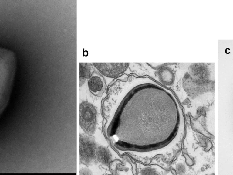
The discovery crystallized around the intricate host-parasite interplay between the newly discovered virion and its single-celled photosynthetic host. The target victim of PelV-1 was identified as an uncultured dinoflagellate belonging to the microalgal genus Pelagodinium. As pivotal primary producers in the global marine carbon system, these microeukaryotic flagellates convert atmospheric carbon dioxide into fundamental oceanic biomass. However, subsequent laboratory co-culturing and high-resolution transmission electron microscopy of the isolated viral particles stunned the scientific community: the physical morphology of the pathogen upended standard paradigms of structural virology.
The virion architecture consists of an icosahedral protein capsid roughly 200 nanometers across, terminating in a gigantic, flexible, thread-like filament. Precision nanoscale measurements revealed that this ultra-slender tail, measuring a mere 30 nanometers in diameter, extends outward to an astonishing 2.3 micrometers (2,300 nanometers) in length. To contextualize this scale, the viral tail is more than ten times longer than the capsid itself, comfortably exceeding the physical body length of typical rod-shaped marine bacteria such as Escherichia coli. PelV-1 instantaneously eclipsed all documented structural records across the global virosphere, easily surpassing long-tailed bacteriophages and outstripping the previous record-holder, South America’s Tupanvirus.
The emergence of PelV-1 adds a dramatic new chapter to the expanding catalogue of giant viral discoveries that challenge classical definitions of biological entities. These ongoing explorations share deep thematic synergy with investigations into atypical viral genomic architectures and marine microbial dynamics, affirming that the marine realm conceals evolutionary adaptations that dissolve historical scientific orthodoxies.
Nanoscale Architecture: The Micron Tail and the Stargate Injection Core
The physical anatomy of the PelV-1 virion constitutes a mechanical masterpiece of evolutionary biological engineering. Comprehensive cryo-electron tomography unveiled a pronounced bipolar morphological organization. While one vertex of the icosahedral capsid anchors the extraordinarily long 2.3-micrometer flexible filament, the diametrically opposed pole features a specialized protein protrusion emerging from a five-fold star-shaped portal. Known in giant virus literature as a “stargate,” this specialized seal functions as an iris-like structural aperture through which the virion ejects its genomic payload directly into the host cytoplasm.
The adaptive evolutionary utility of such an exorbitant tail has generated intense scientific inquiry across biophysics and marine ecology. Unlike the rigid, contractile sheaths characteristic of tailed bacteriophages in the family Myoviridae, the tail of PelV-1 exhibits fluid lateral flexibility. Dr. Christopher Schvarcz, co-author of the landmark description, hypothesized that the tail operates as a hydrodynamic harpoon and sensory grappling antenna. In the vast oligotrophic waters of the open Pacific, where planktonic host cells are widely dispersed, the statistical likelihood of an unassisted 200-nanometer capsid colliding directly with a microalgal target is vanishingly small. The multi-micrometer tail expands the virion’s hydrodynamic capture cross-section by orders of magnitude.
Upon drifting into physical contact with a Pelagodinium cell surface, the tail initiates an intricate adhesive attachment cascade. The flexible filament wraps around the microalga’s thecal plates or flagellar apparatus like a microscopic grappling line, mechanically reeling the bulky capsid flush against the host plasma membrane. Once securely docked, the stargate pentamer unseals, forging a continuous hydrophobic channel that facilitates the rapid translocation of viral DNA into the algal cell interior.
Furthermore, transmission electron micrographs revealed an intricate network of exterior surface fibers and lipid membranes adorning the outer capsid shell of PelV-1. This organic mantle provides essential biophysical resilience, buffering the viral particle against severe osmotic shocks in hypersaline marine waters while shielding sensitive viral core enzymes from photodegradation under relentless solar ultraviolet radiation.
The virion filament extends up to 2300 nanometers, surpassing bacteriophages and all known viral appendages documented across the biosphere.
Double-stranded viral DNA encodes hundreds of proteins, including autonomous enzymes for carbohydrate, lipid, and amino acid metabolic pathways.
PelV-1 harbors functional rhodopsin and photosynthetic antenna genes to siphon solar energy for virion assembly independently of host regulation.
Opposite the micro-tail lies a five-lobed stargate opening where the lipid-encased capsid unseals to inject its genomic payload into host cytoplasm.
Genomic Colossus: Auxiliary Metabolic Repertoires and Light-Harvesting Proteins
While the macromolecular morphology of PelV-1 captured immediate attention, the sequencing of its genomic architecture initiated an even deeper paradigm shift. The viral genome consists of a massive linear double-stranded DNA molecule approximately 459 kilobases (kb) in length, encoding more than four hundred predicted open reading frames. In comparison, human influenza A genomes span approximately 13 kilobases and HIV fewer than 10. The genetic repertoire of PelV-1 dwarfs that of numerous free-living cellular endosymbionts, including Carsonella ruddii and Mycoplasma genitalium.
Phylogenomic reconstructions firmly nest PelV-1 within the family Mesomimiviridae, a foundational lineage within the phylum Nucleocytoviricota (nucleocytoplasmic large DNA viruses, or NCLDVs). However, the defining revelation of the genomic analysis was the extraordinary abundance of auxiliary metabolic genes (AMGs). Rather than behaving as an obligate genomic scavenger utterly dependent on host cellular pathways, PelV-1 delivers an extensive suite of metabolic software capable of reprogramming the entire host physiology.
The viral chromosome houses intact enzymatic cassettes dedicated to central carbohydrate processing, lipid metabolism, specialized amino acid breakdown, and specific transmembrane nutrient transporters including aquaporins and sugar permeases. Most astonishingly, bioinformatic screening identified fully functional genes for light-harvesting complex proteins and viral microbial rhodopsins. Upon gaining entry into the Pelagodinium host, PelV-1 synthesizes these photoactive proteins to harvest ambient solar radiation directly, driving proton-motive gradients across membranes to furnish abundant metabolic ATP for virion synthesis even after host photosynthesis is disabled.
Virologist Dr. Alexander Culley emphasizes that this genetic autonomy metamorphoses the infected microalga into a novel biological hybrid termed a “virocell.” The host cell ceases to function as an independent photosynthetic dinoflagellate, transforming into an algorithmically directed viral synthesis bioplatform governed with industrial precision by the PelV-1 genome.
«The 2.3-micrometer appendage of PelV-1 is physically larger than many free-living marine bacteria. We have historically conceptualized viruses as inert, sub-micron geometric capsules housing minimal genetic instructions; however, PelV-1, with its autonomous metabolic machinery and gargantuan filaments, operates far more like an predatory cellular agent.»
Dr. Andrian Gajigan, Oceanographer and Molecular Microbiologist at the University of Hawaiʻi at Mānoa
The Oceanic Viral Shunt: PelV-1 Driving Global Marine Carbon Sequestration
The discovery of PelV-1 carries profound ecological ramifications extending far beyond taxonomic curiosities, exerting direct influence over global climatology and marine biogeochemical cycles. Marine systems ecology relies heavily upon the paradigm of the viral shunt—a fundamental planetary mechanism whereby marine phages and giant viruses lyse an estimated 20% to 40% of all prokaryotic and eukaryotic planktonic biomass daily. This incessant cellular lysis short-circuits traditional trophic pathways, diverting organic carbon away from macro-grazers and returning petagrams of carbon to the dissolved organic matter (DOM) pool.
Viruses of the family Mesomimiviridae targeting Pelagodinium serve as supreme ecological arbiters of marine algal population blooms. During favorable subtropical upwelling conditions, photosynthetic microalgae proliferate explosively across thousands of square kilometers. Yet this rapid biomass accumulation triggers synchronized, epidemic propagation of PelV-1. The resultant mass lysis of millions of algal cells releases immense volumes of intracellular cellular debris, structural proteins, and polysaccharides directly into the water column.
These sticky, lysed cellular remnants aggregate into macroscopic organic flocs designated by marine scientists as “marine snow.” Driven by gravity, these dense aggregations sink inexorably through the twilight bathypelagic layers into the abyssal ocean interior, effectively sequestering atmospheric carbon within seafloor sediments for geological timescales. Consequently, this giant virus wielding an unprecedented tail operates as a microscopic planetary climate regulator, governing the capacity of the Pacific Ocean to buffer global anthropogenic greenhouse gas emissions.
Crucially, deep metagenomic sequencing from Station ALOHA revealed the presence of a co-occurring sister lineage dubbed co-PelV, possessing an even larger 504-kilobase genome. This confirms that oligotrophic ocean gyres sustain complex, multi-tiered viral consortia that coordinate primary productivity and carbon pump mechanics across vast oceanic regimes.
- Isolated from oligotrophic surface waters north of Oahu, Hawaii, at the long-term ocean research Station ALOHA.
- Exclusively infects single-celled photosynthetic Pelagodinium dinoflagellates responsible for critical marine primary production.
- Taxonomically classified within the nucleocytoplasmic large DNA virus (NCLDV) family Mesomimiviridae.
- The 2.3-micrometer tail surpasses the former record holder, the Brazilian Tupanvirus, by more than a full micrometer in length.
Dissolving the Living Boundary: Philosophic and Evolutionary Status of Giant Virions
The characterization of PelV-1, armed with its 2.3-micrometer locomotive appendage and extensive metabolic machinery, reinvigorates an unresolved foundational debate: what fundamentally constitutes living matter? Twentieth-century biological dogma established an inflexible barrier segregating non-living acellular viral replicators from true cellular organisms, defined by the absence of endogenous metabolism, inability to generate autonomous energy, and absolute dependence on foreign ribosomal translation. Yet giant NCLDV virions methodically dismantle every pillar of this demarcation.
When an entity boasts a genome larger than hundreds of bacterial taxa, manufactures its own photoactive rhodopsins to synthesize energy, and produces metabolic enzymes across diverse pathways, designating it merely as an inert nucleoprotein package represents untenable reductionism. Professor Grieg Steward contends that giant viruses of the PelV-1 caliber represent an ancient, co-equal evolutionary lineage that has co-evolved alongside Archaea, Bacteria, and Eukarya since the dawn of terrestrial life.
Intense phylogenetic investigation centers upon the evolutionary genesis of these colossal entities. Under the reductive degradation hypothesis, ancestral precursors of modern Mesomimiviridae were once autonomous, free-living proto-cellular lineages that adopted obligate intracellular parasitism, shedding bulky ribosomal translation machinery while preserving complex metabolic cassettes. Conversely, the gene accretion hypothesis posits that an ancestral small virus acted as an evolutionary sponge, progressively accumulating functional host genes across eons of horizontal gene transfer.
Ultimately, PelV-1 demonstrates that nature recognizes no rigid anthropogenic taxonomies. This dweller of the open Pacific thrives within the ambiguous borderland of the biosphere—bridging inert crystalline chemistry and complex cellular self-determination. The study of PelV-1 compels biological science to abandon binary definitions of life, acknowledging instead a seamless continuum of self-organizing matter whose most astounding oceanic expression is embodied by this giant, long-tailed oceanic viral titan. Also read our related investigative feature on atypical viral genomic architectures and marine microbial dynamics.