

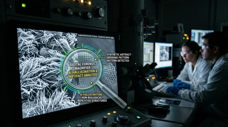

In a seismic reckoning for scientific imaging, the Nikon Small World cilia video disqualified generative AI controversy has shocked cell biologists worldwide. A high-profile microscopic entry that claimed First Prize in the 2026 Nikon Small World in Motion competition — purportedly displaying ultra-high-resolution dynamic beating of human respiratory epithelial cilia in Primary Ciliary Dyskinesia (PCD) — has been stripped of its crown. An international coalition of cilia biologists and biophysicists demonstrated that the captivating footage was not genuine microscopy, but a synthetic hallucination generated by artificial intelligence that fundamentally violated the universal 9+2 microtubule axoneme architecture of eukaryotic life.
Prestige Shattered: How an AI Hallucination Fooled Nikon Small World Judges
For over fifty years, the Nikon Small World and Nikon Small World in Motion competitions have stood as the premier international benchmark for bioimaging excellence. Celebrating the intersection of rigorous optical microscopy and artistic marvel, the awards celebrate authenticated captures of microscopic phenomena submitted by leading university labs and biomedical researchers.
The 2026 Small World in Motion first-place prize was awarded to a hypnotic sequence depicting airway epithelial cilia from a patient suffering from Primary Ciliary Dyskinesia (PCD). The judges were enamored by the crystal-clear contrast, lack of typical optical noise, and cinematic depth-of-field across the moving cellular carpet.
However, celebration turned to condemnation within hours of the public broadcast. Cell biologists specializing in motile ciliopathies immediately identified glaring anatomical anomalies that could not exist in real biological specimens.
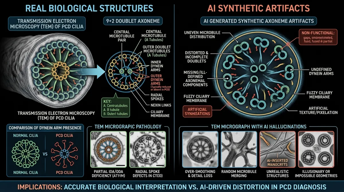
Cellular Ultrastructure: The Non-Negotiable 9+2 Axoneme Architecture
At the heart of all eukaryotic motile cilia lies the axoneme — a highly conserved macromolecular engine. Cross-sectional transmission electron microscopy invariably reveals a strict 9+2 geometric layout: nine outer microtubule doublets interconnected by nexin links and inner/outer dynein arms surrounding a central microtubule pair.
The ATP-driven sliding of dynein motor arms generates the characteristic biphasic stroke: a rapid, planar effective stroke followed by a curved recovery stroke hugging the cell surface. In patients with Primary Ciliary Dyskinesia, mutations in dynein genes like DNAH5 paralyze the cilia or produce stiff, uncoordinated micro-tremors.
In stark contrast, the contest-winning footage showed cilia waving in synchronous, sinusoidal ribbons. High-magnification frame-by-frame scrutiny revealed that individual cilia bifurcated, merged into neighboring fibers, and dematerialized into the fluid background — telltale hallmarks of diffusion-based generative video models interpolating pixels.
Nikon Small World in Motion stripped its top prize after experts proved the video utilized generative AI interpolation.
The generated cilia lacked essential microtubule doublets and dynein motor arms dictated by eukaryotic biology.
Fluid mechanics simulations proved cilia movements violated hydrodynamic laws governing microscopic cellular viscous drag.
New contest standards enforce cryptographic verification of native camera sensor files and microscope optics logs.
Biophysical Sleuthing: Exposing Hydrodynamic Violations
A forensics review led by cilia researchers subjected the video to hydrodynamic simulation. At the cellular microscale, fluid physics is governed by extremely low Reynolds numbers ($Re \ll 1$), where viscous drag overwhelms inertial forces. In this regime, water feels as thick as molten asphalt; cilia cannot exhibit inertial momentum or coasting oscillation waves.
The AI-generated video depicted buoyant, frictionless wave motion completely incompatible with fluid mechanics at the micron scale. Moreover, phase-contrast Fourier transforms revealed a total lack of native optical diffraction rings, displaying smooth computer-generated polygon shading.
Presented with this decisive biophysical dossier, the Nikon Small World committee launched an emergency inquiry. The creator admitted to utilizing AI diffusion upscaling and synthetic generative video interpolation to fabricate missing temporal data, corrupting the scientific authenticity of the footage.
«To a casual observer, the footage appeared mesmerizingly fluid and sharp, but to any respiratory biologist, it was instantly preposterous. Cilia do not bend like rubbery seaweed, nor do their microtubule doublets dissolve into empty space. Generative AI created an aesthetically pleasing rendering while completely trampling basic cell biology.»
Matthias Salathe, Professor of Internal Medicine and international authority on airway epithelial cilia
Scientific Integrity Under Threat: Why Hallucinated Imaging Matters
Nikon acted decisively by revoking the award, purging all entries from official media, and issuing an apology to the scientific community. It marks the first disqualification in the half-century history of the competition driven by generative artificial intelligence.
The incident carries profound ramifications far beyond photography contests. In biomedical science, imaging is not decorative; it is empirical evidence. If generative models hallucinate nonexistent structural phenotypes, pharmaceutical developers could misinterpret drug efficacy when designing targeted therapeutics for rare respiratory syndromes.
Scientific visualization requires uncompromising physical truth, mirroring standards in optical microscopy and bioimaging, where every pixel represents photons interacting with genuine biological matter rather than synthetic probabilities.
- The entry claimed to show human respiratory epithelial cilia beating in Primary Ciliary Dyskinesia (PCD).
- Specialist cilia biologists exposed artificial bifurcations and frictionless fluid dynamics generated by AI.
- Generative diffusion models hallucinated impossible biological structures to simulate high framerates.
- The scandal prompted international imaging competitions to mandate unedited RAW sensor validation.
Enforcing Rigorous Verification: The RAW Telemetry Protocol
In the wake of the scandal, bioimaging competitions worldwide are overhauling submission protocols. Moving forward, finalists must provide native camera RAW sequences, complete instrument metadata (laser wavelength, objective numerical aperture, pinhole settings), and raw calibration bead controls.
Microscope manufacturers are developing hardware-level cryptographic watermarks that sign sensor captures at the focal plane, preventing generative tampering. Full reportage on the scientific fallout is documented on IFLScience.
Nikon Small World 2026 will be remembered as the moment the scientific world drew an immutable line: the beauty of the microscopic universe has value only when it reflects reality.