



For generations, classical dietetics and human physiology categorized adipose tissue solely as an inert energy storage depot—a passive reservoir of excess triglycerides associated with systemic inflammation and metabolic disorders. However, that description pertains exclusively to white adipose tissue. A profound biological revolution unfolded when functional brown adipose tissue (BAT) was identified in adult humans. Rather than conserving energy, these specialized adipocytes operate as microscopic cellular incinerators: they avidly extract circulating glucose and fatty acids from the bloodstream and dissipate them as pure thermal energy through the unique action of uncoupling protein 1 (UCP1, or thermogenin).
White, Brown, and Beige: The Anatomy of a Metabolic Marvel
The human body harbors three distinct varieties of adipocytes. White adipose tissue (WAT) consists of spherical cells containing a single unilocular lipid droplet engineered for long-term caloric storage. In stark contrast, brown adipose tissue (BAT) features multilocular architecture: its cells are packed with numerous minute lipid droplets and a dense concentration of iron-rich mitochondria containing pigmented cytochromes, which bestow the tissue with its characteristic reddish-brown hue.
Historically, medical dogma asserted that BAT was confined to human infants—who require thermal protection due to their high surface-area-to-mass ratio—and hibernating mammals. Only with the advent of 18F-fluorodeoxyglucose positron emission tomography (PET-CT) in the late 2000s did researchers conclusively establish that adult humans retain metabolically active BAT depots localized in the supraclavicular, cervical, paravertebral, and suprarenal anatomical regions.
Furthermore, scientists unraveled the process of “browning” (or beiging), wherein prolonged physiological stimulation induces white adipocyte precursors to transdifferentiate into beige (or “brite”) adipocytes, acquiring thermogenic mitochondrial machinery and UCP1 expression capacity.
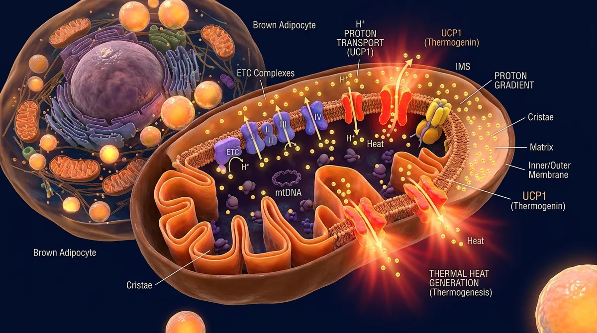
The Biochemistry of Uncoupling: How UCP1 Hacks the Mitochondria
Under standard cellular respiration conditions, the mitochondrial electron transport chain oxidizes metabolic substrates, pumping hydrogen protons across the inner mitochondrial membrane into the intermembrane space. This creates an electrochemical proton motive force, compelling protons to re-enter the mitochondrial matrix exclusively through the ATP synthase rotary complex, thereby generating cellular energy.
In brown adipocytes, this fundamental bioenergetic mechanism is fundamentally decoupled. Their inner mitochondrial membrane is heavily enriched with uncoupling protein 1 (UCP1). UCP1 forms a specialized proton conductance pathway that bypasses ATP synthase entirely. Protons collapse back into the matrix down their electrochemical gradient, releasing stored potential energy purely as heat.
This non-shivering thermogenesis allows brown adipocytes to function at maximum oxidative capacity without accumulating excessive ATP. In doing so, they consume vast quantities of glucose, circulating free fatty acids, and branched-chain amino acids.
Brown fat does not store excess calories; it incinerates lipids due to an extreme concentration of iron-rich mitochondria.
Thermogenin short-circuits the proton gradient around ATP synthase, dissipating all energy directly as thermal heat.
Cold exposure triggers sympathetic beta-3 adrenergic receptors, driving instant lipolysis and UCP1 derepression.
Active BAT consumes substantial glucose and triglycerides, providing powerful protection against diabetes and plaque.
The Signaling Cascade: Noradrenaline and Beta-3 Adrenergic Receptors
The primary physiological trigger for brown fat thermogenesis is environmental cold exposure. Cutaneous thermoreceptors detect ambient temperature drops and transmit afferent neural signals to the preoptic area of the hypothalamus. The hypothalamus subsequently activates the sympathetic nervous system (SNS), dispatching efferent signals directly to perivascular BAT depots.
Postganglionic sympathetic terminals release the neurotransmitter noradrenaline, which binds selectively to beta-3 adrenergic receptors situated on the brown adipocyte plasma membrane. This interaction stimulates adenylate cyclase, elevating intracellular cyclic AMP (cAMP) and activating protein kinase A (PKA).
PKA rapidly phosphorylates hormone-sensitive lipase, initiating lipolysis within the multilocular lipid droplets. The released free fatty acids fulfill a vital dual role: they serve as immediate oxidative substrates for mitochondrial beta-oxidation and directly allosterically activate UCP1 by relieving purine nucleotide inhibition, initiating full thermogenic capacity within minutes.
«Brown adipose tissue is a marvelous evolutionary thermogenic engine. While typical somatic cells meticulously conserve chemical energy within the high-energy phosphate bonds of ATP, brown adipocyte mitochondria deliberately short-circuit this pathway via UCP1. The electrochemical proton gradient is discharged directly as heat. Activating even a few grams of brown fat can elevate daily basal expenditure by hundreds of kilocalories while substantially improving systemic insulin sensitivity.»
Dr. Bruce Spiegelman, Professor of Cell Biology and Medicine at Dana-Farber Cancer Institute, Harvard Medical School
Clinical Implications: Counteracting Obesity, Type 2 Diabetes, and Atherosclerosis
Because active brown adipose tissue requires vast biochemical fuel to maintain thermogenesis, its sustained stimulation exerts profound whole-body cardiometabolic benefits. Clinical investigations demonstrate that cold-activated BAT markedly accelerates whole-body glucose clearance through insulin-independent pathways, significantly attenuating insulin resistance in patients with type 2 diabetes.
Additionally, active BAT serves as a metabolic sink for triglyceride-rich lipoproteins and very-low-density lipoproteins (VLDL), protecting hepatic tissue from steatosis and slowing the atherosclerotic plaque cascade within arterial walls.
Individuals exhibiting greater brown fat volume display lower circulating inflammatory cytokines, improved lipid profiles, stabilized blood pressure, and higher basal metabolic rates (BMR) even in thermoneutral resting environments.
- Adult humans maintain active brown fat depots primarily in the supraclavicular, cervical, and paravertebral regions.
- Uncoupling protein 1 (UCP1) is directly activated by free fatty acids released during noradrenaline-driven lipolysis.
- Mild ambient cold exposure (16–18°C) stimulates the browning of white adipocytes into highly oxidative beige fat.
- Brown adipose tissue operates as an insulin-independent glucose sink, reversing metabolic dysregulation.
Evidence-Based Protocols: How to Stimulate and Brown Your Adipocytes
Translational endocrinology has outlined actionable, evidence-based regimens to recruit and activate thermogenic fat depots. Mild cold exposure represents the most reliable intervention: daily exposure to temperatures between 16°C and 18°C (60–64°F) for two hours across six weeks expands active BAT volume by 40% to 50% and enhances cold-induced thermogenesis.
Complementary modalities include brief cold water immersion, contrast showers, and high-intensity interval exercise—which stimulates muscle secretion of irisin, a myokine that robustly triggers the browning of subcutaneous white fat. Dietary bioactives such as capsaicin from chili peppers and resveratrol further synergize with adrenergic pathways, offering a multifaceted approach to metabolic optimization.