

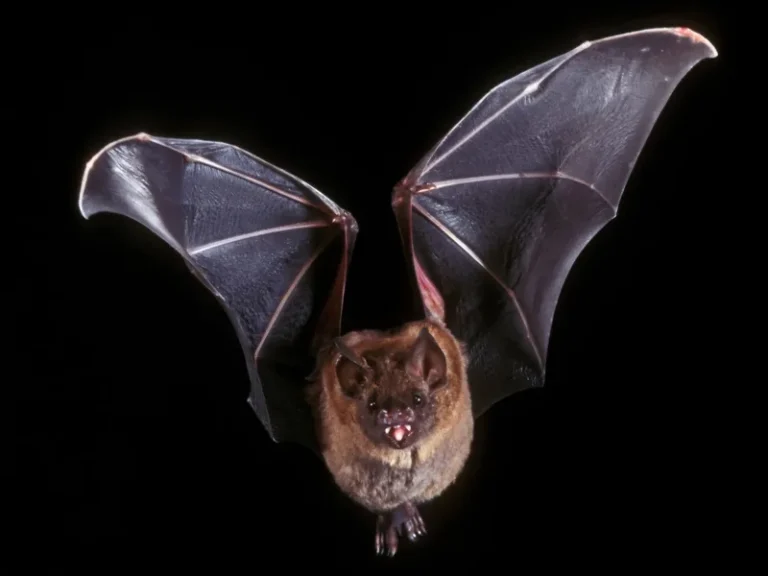

Navigating through pitch-black subterranean caverns or turbid ocean abysses without visual cues represents one of the crowning achievements of convergent evolution. Echolocation biosonar bats dolphins transformed high-frequency acoustic vibrations into a hyper-detailed three-dimensional sensory landscape. By exploring the neurobiology of auditory cortex processing in microbats and odontocetes, researchers have discovered that biological sonars calculate target distance with sub-millimeter precision and decode internal object densities faster than advanced military radars.
Acoustic Physics of Biosonar: Frequency Modulation and Constant Frequency
At the foundation of biological echolocation lies the universal wave mechanics of acoustic reflection at acoustic impedance boundaries. Over millions of years of predatory pressure, mammalian evolution refined this physical principle into two distinct echolocation paradigms: broadband frequency-modulated (FM) chirps and narrowband constant-frequency (CF) transmissions. Each strategy represents an optimal trade-off between target identification and spatial ranging accuracy.
Most vespertilionid bats deploy ultra-short broadband FM sweeps lasting 1 to 3 milliseconds, during which carrier frequency cascades downwards—for instance, sweeping from 100 kHz down to 25 kHz. This broad spectral breadth yields immense information concerning target geometry, surface texture, and angular wing aspect. By analyzing microsecond differences in binaural arrival times and spectral notch filtering, the bat’s auditory cortex constructs a crisp volumetric representation of fluttered moth wings.
In contrast, rhinolophid (horseshoe) bats utilize extended constant-frequency (CF) pulses hovering around 83 kHz, terminated with a brief FM down-sweep. Continuous-frequency emissions are uniquely tuned for detecting prey against cluttered background foliage via the Doppler effect: the rhythmic fluttering of an insect’s wings induces micro-Doppler glints, creating dynamic frequency sidebands that cut through dense leaf echoes.

Anatomical Sound Generators: Laryngeal Specialization and the Dolphin Melon
The biomechanical systems driving biological ultrasound generation are marvels of functional anatomy. In bats, acoustic pulses originate in an ultra-specialized larynx equipped with superfast vocal fold musculature capable of contracting more than 200 times per second. The cricothyroid muscles exhibit the highest mitochondrial and sarcoplasmic reticulum densities known in mammalian physiology, sustaining rapid twitch kinetics without lactic acidosis.
To propagate acoustic energy into free space, bats either use an open mouth acting as an acoustic horn or project sound through intricate fleshy nasal structures known as noseleaves. These nasal folds operate as spatial diffraction gratings and acoustic resonators, sharpening the main transmission beam while suppressing destructive sidelobes that would generate blinding reverberations from nearby foliage.
Cetacean anatomy evolved along a fundamentally divergent path. Toothed whales lack vocal cords altogether. Instead, situated within the nasal passage below the blowhole are paired muscular structures known as the “phonic lips” (monkey lips) coupled to air sacs. By pressurizing air across these valve-like margins, the animal forces the lips into violent cyclic closures, generating explosive mechanical transients.
Auditory Neuroscience: The Acoustic Fovea and Central Delay-Tuned Maps
How does the mammalian central nervous system process an onslaught of overlapping echoes returning with microsecond latency differences? In bats, returning sound waves are collected by large, mobile pinnae featuring an exaggerated tragus. The structural convolutions of the pinna act as an angle-dependent spectral notch filter: vertical elevation is computed instantaneously from characteristic spectral notches without requiring head tilting.
Within the cochlea of horseshoe bats lies an evolutionary marvel: the acoustic fovea. This represents a dramatic physical hypertrophy of the basilar membrane, where over 20% of all primary hair cells are tuned to a razor-thin band spanning barely 2 kHz around the resting frequency (83 kHz). Functioning exactly like the retinal fovea centralis of the eye, this cochlear specialization yields acute frequency discrimination, detecting echo Doppler shifts as small as 0.05%.
In dolphins, the external ear canals are vestigial and filled with dense cellular debris. Sound reception underwater occurs primarily through the lower jaw, which houses an acoustic lipid channel known as the intramandibular fat pad. Acoustic waves in the surrounding water penetrate the thin acoustic window of the mandibular bone with minimal impedance loss, propagating along the lipid tract directly to the isolated tympanoperiotic complex of the middle and inner ear.
Within the brainstem, echo analysis is coordinated by the inferior colliculus and the primary auditory cortex. Neurophysiologists have identified delay-tuned combination-sensitive neurons: these cells fire selectively only when an echo follows an emitted pulse with an exact temporal delay (e.g., precisely 2.5 milliseconds). Arranged in columnar arrays, these neurons form an orderly topographic computational map of external target distance.
The echolocating brain does not merely register sound echoes — it synthesizes a coherent spatial representation with temporal precision reaching the tens of nanoseconds.
Dr. James Simmons, Professor of Neuroscience and Bioacoustics at Brown University
Molecular Convergence: Parallel Amino Acid Substitutions in Prestin
One of the most profound revelations of modern molecular phylogenetics was the discovery of extensive genetic convergence between bats and cetaceans. Although bats and dolphins diverged from their common placental ancestor more than 85 million years ago, the biophysical necessity of detecting acoustic signals above 100 kHz compelled nature to arrive at nearly identical genetic adaptations.
Central to high-frequency auditory mechanics is prestin (SLC26A5), a transmembrane motor protein residing within the lateral membranes of cochlear outer hair cells. Prestin functions as an ultra-rapid piezoelectric actuator: driven by changes in membrane potential, it undergoes conformational length changes within microseconds, mechanically amplifying basilar membrane vibrations by several orders of magnitude.
Comparative sequence analysis revealed a stunning coincidence: 14 identical amino acid substitutions in prestin were shared between echolocating odontocetes and horseshoe bats. In standard terrestrial mammals, prestin-driven amplification collapses at frequencies above 30 kHz due to internal viscous damping. However, these convergent substitutions altered the protein’s charge-displacement mechanics, enabling high-gain cochlear amplification up to 160 kHz.
Biomimetic Frontiers: From Animal Sonar to Medical Ultrasound and Drone Autonomy
Decoding natural biosonar has transcended comparative physiology to become a major driver of modern robotics, biomimetic acoustics, and medical imaging. For decades, naval sonar arrays struggled to replicate the dolphin’s ability to discriminate between hollow and solid metallic cylinders buried under seabed sediment—a computational feat odontocetes execute in fractions of a second.
Next-generation bio-inspired sonar systems modeled on mammalian auditory pathways abandon rigid square-pulse transmissions in favor of dynamic frequency sweeps and adaptive beam steering. This biomimetic approach reduces sonar energy consumption aboard autonomous underwater vehicles (AUVs) by more than 80%, enabling prolonged subsea reconnaissance missions.
In aerial robotics, bio-inspired ultrasonic sensor arrays combined with Doppler-compensation processors enable micro-drones to navigate through dense forest undergrowth at speeds exceeding 40 km/h in absolute darkness. By analyzing multi-path echo interference from tangled branches, neuromorphic processors plot collision-free flight trajectories within milliseconds.