

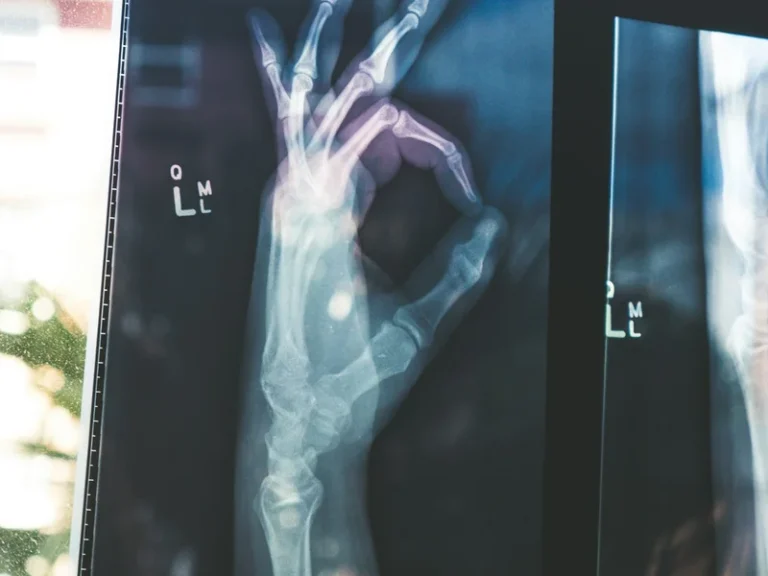

Human biological aging was long regarded as an unavoidable accumulation of random cellular mutations and mechanical wear-and-tear. However, pioneering breakthroughs in biogerontology and mucosal immunology have exposed a primary systemic driver: the phenomenon of inflammaging. Chronic inflammation inflammaging gut permeability aging links gut dysbiosis directly to degenerative diseases. When the microscopic single-cell epithelial lining of the intestine loses its tight junction integrity, bacterial endotoxins seep into the bloodstream, igniting a persistent immune blaze that damages arterial vessels and destroys brain synapses.
Anatomy of the Mucosal Barrier: A 30-Micrometer Defensive Monolayer
The human gastrointestinal tract constitutes the body’s largest interface with the external environment: when accounting for mucosal plicae, villi, and microvillar brush borders, its absorptive surface area exceeds 32 square meters. This vast territory houses over 70% of the entire human immune system and harbors an ecological community of over 40 trillion microorganisms. Remarkably, this bustling micro-world is separated from our sterile internal bloodstream by a single layer of epithelial cells barely 30 micrometers thick.
Under healthy physiological conditions, adjacent enterocytes and colonocytes are joined by multiprotein complexes known as tight junctions (TJ). Composed of transmembrane claudins, occludin, and cytosolic scaffolding adaptors (zonula occludens ZO-1 and ZO-2), these complexes act as dynamic nanometric gatekeepers: they permit passive paracellular transport of water, electrolytes, and vital nutrients while blocking intact bacteria, dietary macromolecules, and microbial endotoxins.
Shielding this cellular monolayer is a thick, hydrated mucus blanket secreted by specialized goblet cells. The inner layer of MUC2 mucin is dense and essentially impervious to bacteria, while the looser outer layer provides a rich ecological habitat for symbiotic commensals. Secretory immunoglobulin A (sIgA) dimers continually neutralize invasive flagellated microbes before they can engage epithelial apical membranes.
However, the modern lifestyle—defined by ultra-processed foods, chronic psychological stress, antibiotic overutilization, and non-steroidal anti-inflammatory drugs (NSAIDs)—compromises this defensive network. Depleted mucus layers and uncoupled tight junctions produce pathological intestinal hyperpermeability, popularly known as “leaky gut syndrome”.
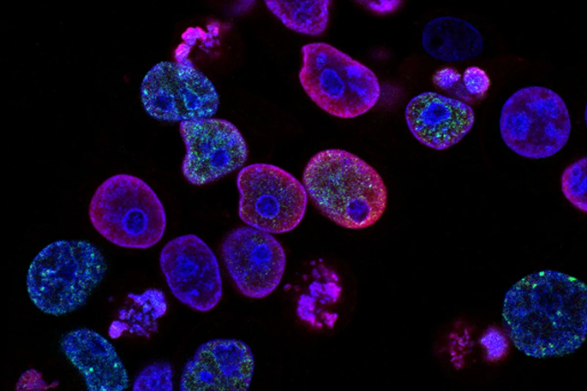
Molecular Subversion: Zonulin, LPS Translocation, and Metabolic Endotoxemia
The primary physiological modulator of paracellular permeability is the protein zonulin, discovered by Harvard mucosal immunologist Dr. Alessio Fasano. In response to gliadin peptides from gluten or colonization by opportunistic bacteria, enterocytes hypersecrete zonulin. Upon binding epidermal growth factor (EGFR) and protease-activated (PAR2) receptors, zonulin activates a phospholipase C cascade that polymerizes actin stress fibers, physically wrenching tight junctions apart.
Through these widened paracellular apertures escapes the primary instigator of modern chronic disease: lipopolysaccharide (LPS), a glycolipid constituent of gram-negative bacterial outer envelopes. While harmless within the intestinal lumen, LPS molecules that leak into the lamina propria and capillary beds provoke a profound immune response.
Once in systemic circulation, LPS binds lipopolysaccharide-binding protein (LBP) and engages Toll-like receptor 4 (TLR4) on circulating monocytes and tissue-resident macrophages. For the mammalian innate immune system, TLR4 activation mimics severe bacterial sepsis.
Downstream, the nuclear factor-kappa B (NF-kB) transcriptional pathway mobilizes, churning out an unrelenting stream of pro-inflammatory cytokines: interleukin-1-beta (IL-1β), interleukin-6 (IL-6), and tumor necrosis factor-alpha (TNF-α). Because endotoxin leakage is chronic rather than acute, it establishes low-grade metabolic endotoxemia—the physiological cornerstone of systemic inflammaging.
Lipopolysaccharides (LPS) shed from gram-negative bacterial outer walls bypass degraded tight junctions into the portal vein, stimulating TLR4 receptors and inducing systemic endotoxemia.
Excess mucosal zonulin secretion triggers actin cytoskeleton contraction, disassembling occludin and claudin protein complexes to convert selective barriers into leaky sieves.
Circulating pro-inflammatory cytokines IL-6 and TNF-alpha disrupt the blood-brain barrier, converting quiescent brain microglia into destructive neurotoxic M1 phenotypes.
Microbiome-derived butyrate supplies metabolic fuel for colonocytes, stimulating mucin synthesis and upregulating tight junction protein assembly to mitigate aging.
Cardiovascular Vulnerability: Endotoxin-Induced Atherosclerosis and Plaque Rupture
Low-grade chronic inflammation is a decisive driver of cardiovascular morbidity, including myocardial infarction and ischemic stroke. Classical lipid-centric paradigms fail to explain why more than 50% of acute coronary events occur in individuals possessing normal low-density lipoprotein (LDL) cholesterol profiles. Endothelial immunology provides the missing mechanistic link.
The vascular endothelium is coated by a protective carbohydrate meshwork called the endothelial glycocalyx. Circulating LPS endotoxins and systemic IL-6 enzymatically degrade this glycocalyx shield, activating endothelial cells to express vascular cell adhesion molecules (VCAM-1 and ICAM-1) that tether circulating blood monocytes.
Tethered monocytes extravasate into the subendothelial intima, differentiate into macrophages, and engulf oxidized LDL particles to form lipid-laden foam cells—the genesis of an atherosclerotic plaque. In an environment devoid of systemic inflammation, these plaques remain stable beneath protective collagen fibrous caps for decades.
However, sustained endotoxemia prompts intralesional macrophages to release matrix metalloproteinases (notably MMP-9), which digest the structural collagen cap. The fibrous cap weakens, exposing thrombogenic core lipids to coagulation factors in the blood. Platelets aggregate within seconds, producing an occlusive coronary thrombus that precipitates an acute heart attack.
«The intestinal barrier separates our internal physiology from trillions of resident microbes with just a single monolayer of epithelial cells. Compromising this boundary turns our own microbiome into a persistent inflammatory engine.»
Dr. Alessio Fasano, Professor of Pediatrics at Harvard Medical School
The Gut-Brain Axis: Blood-Brain Barrier Breakdown and Microglial Activation
The repercussions of mucosal barrier compromise are equally devastating for cerebral longevity. Communication along the gut-brain axis occurs via three interconnected routes: the vagus nerve, enteric neuroendocrine signaling, and humoral systemic circulation.
The cerebral parenchyma is insulated from systemic toxins by the blood-brain barrier (BBB), maintained by tight junctions between capillary endothelial cells and astrocytic end-feet. Unfortunately, circulating LPS and TNF-α downregulate brain endothelial occludin and claudin-5 in the exact same manner as in the gut. The blood-brain barrier becomes porous.
Crossing into the central nervous system, LPS binds TLR4 complexes on microglia—the brain’s resident phagocytes. Under basal conditions, ramified microglia (M2 phenotype) nurture synapses, promote neurogenesis, and clear metabolic debris. Exposure to circulating endotoxins shifts microglia into an ameboid, neurotoxic M1 phenotype.
M1 microglia flood neural pathways with reactive oxygen species (ROS), nitric oxide, and neurotoxic cytokines, producing sustained neuroinflammation. In this hostile microenvironment, hippocampal dendritic spines wither, impairing spatial memory, driving clinical depression, and accelerating the toxic aggregation of amyloid-beta and tau proteins characteristic of Alzheimer’s disease.
- High-sensitivity C-reactive protein (hs-CRP) above 1.5 mg/L signals smoldering vascular inflammation.
- Circulating LPS endotoxin strips the coronary endothelial glycocalyx, inciting unstable plaques.
- Senescent cells (SASP) continuously secrete inflammatory chemokines that degrade tissue matrix.
- Loss of microbiome species diversity diminishes endogenous butyrate synthesis by over 60%.
Therapeutic Restoration: Microbiome Pharmacology for Longevity
Can this insidious inflammatory cascade be reversed to decelerate biological aging? Advances in microbiome therapeutics and functional gastroenterology demonstrate that resolving mucosal hyperpermeability is fully achievable through targeted nutritional and pharmacological interventions.
Foremost among mucosal healing agents are short-chain fatty acids (SCFAs), specifically butyrate, propionate, and acetate. Butyrate serves as the primary energetic substrate for colonocytes, generating over 70% of their cellular ATP via mitochondrial beta-oxidation. Beyond energy production, butyrate upregulates tight junction protein transcription and stimulates protective mucin secretion.
To boost endogenous butyrate synthesis, dietary regimens must supply prebiotic soluble fibers: inulin, chicory fructooligosaccharides, resistant starches, and oat beta-glucans. Clinical trials also validate L-glutamine supplementation—a conditionally essential amino acid that fuels enterocyte proliferation—and zinc-carnosine, which stabilizes mucosal membranes and accelerates mucosal repair.
Authentic systemic rejuvenation begins not with cosmetic therapies, but deep within the intestinal crypts. By fortifying the gut epithelial monolayer and nurturing keystone commensal microbes (such as Akkermansia muciniphila and Faecalibacterium prausnitzii), individuals can silence metabolic endotoxemia at its root, safeguarding the heart, vasculature, and brain across decades of vibrant longevity. Also read our related investigative feature on microbiome-immune interactions and neurobiological systems.