



In a breathtaking convergence of artificial intelligence and cognitive neuroscience, researchers have unveiled an unprecedented deep learning architecture: ai decodes brain activity into pictures using non-invasive functional magnetic resonance imaging (fMRI) data, reconstructing what a person sees with remarkable fidelity. The new model, named Brain-IT, accomplishes an elusive dual feat—transforming complex neural activity into high-resolution images while reversibly predicting how the living human brain will respond to any digital picture.
From Voxels to Pixels: Overcoming Historical Roadblocks in Neural Decoding
Peering directly into the visual theater of the human mind has long stood as one of science’s greatest ambitions. Past attempts to interpret functional MRI data faced severe physiological bottlenecks: scanners measure blood-oxygen-level-dependent (BOLD) hemodynamics rather than the millisecond electrical firings of individual neurons. The fundamental scanning unit is a “voxel”—a tiny 3D cube of brain tissue just millimeters across that houses hundreds of thousands of neurons and millions of synapses.

Historically, machine learning decoders were notoriously brittle, requiring custom, exhaustive training sessions for each specific subject. If a volunteer stepped out of the scanner or if testing migrated to a machine with a different magnetic field strength (such as 3 Tesla versus 7 Tesla), the model’s predictive power collapsed. A research team at the Weizmann Institute of Science in Israel, led by doctoral candidate Roman Beliy and Professor Michal Irani, has now shattered this constraint.
Their framework unites two complementary architectures: the Universal Brain Encoder and Brain-IT (Brain Image Transformation). Much like how international decoding of complex neural circuitry and brain connectome structures reveals shared wiring principles across species, the Weizmann architecture taps into invariant organizational rules governing the human visual cortex.
The Dual-Stream Architecture: Separating Spatial Structure from Semantic Meaning
Presented at the prestigious ICLR conference and documented in an extensive preprint on arXiv, Brain-IT’s core breakthrough lies in assigning a learned vector profile to every voxel in the visual cortex. Each microscopic volume learns to report what visual features it cares about—edges, orientation, surface textures, or high-level abstractions like human faces and animal silhouettes.
The algorithm clusters thousands of voxels spanning early visual areas (V1, V2, V4) and the lateral occipital complex into 128 functional groups. Utilizing cross-attention mechanisms, these clusters share contextual clues, filtering out cardiac and respiratory hemodynamic noise.
Brain-IT outputs two parallel predictions: 1) Structural information (the spatial layout, geometry, and boundary outlines of the scene); 2) Semantic information (conceptual meaning, such as “a baseball batter on a field” or “a dog sitting in grass”). These predictions are fed into a pre-trained generative diffusion model. Rather than initiating generation from pure random noise, the diffusion model crystallizes pixels guided directly by the brain’s structural skeleton.
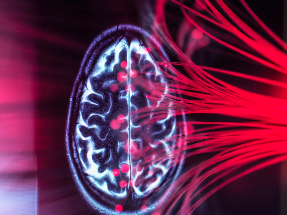
«We are not reading people’s thoughts. The images we do manage to recover from fMRI require people’s cooperation and full intended collaboration. It reflects active visual perception, not inner mental musings.»
Prof. Michal Irani, Study Co-Author, Weizmann Institute of Science
Bidirectional Modeling: Predicting Neural Responses from Arbitrary Images
A remarkable feature of the system is its bidirectional versatility. While Brain-IT reconstructs images from recorded brain scans, the Universal Brain Encoder works in reverse (Image-to-fMRI), predicting the exact voxel activation patterns that will ignite in a human brain when shown any arbitrary photograph or graphic.
This bidirectional bridge offers profound implications for computational neuroscience: researchers can rigorously test hypotheses regarding sensory cortical hierarchy “in silico,” simulating human neural responses on a server without requiring volunteers to spend grueling hours inside claustrophobic MRI bores.
Furthermore, the shared latent space allows seamless cross-subject generalization. Trained on one cohort of participants, Brain-IT can decode images from the brain of a completely new subject viewing novel visual stimuli, confirming the universal architecture of human visual perception.
The model groups thousands of visual cortex voxels into 128 clusters based on sensory tuning.
Decodes spatial geometry and high-level semantics separately to steer a diffusion generator.
Reconstructs seen pictures from brain scans and predicts brain responses from arbitrary images.
Incapable of decoding abstract thoughts, memories, or dreams without active visual perception.
Ethical Guardrails: Can AI Read Our Private Thoughts and Memories?
The emergence of visual neuro-decoders inevitably provokes public unease regarding dystopian prospects of covert mind reading, involuntary surveillance, or the extraction of private memories. The study’s authors emphasize that biological and technical realities make involuntary thought decoding impossible.
Brain-IT decodes exclusively from the primary visual cortex, which processes real-time sensory inputs arriving directly from the retinas. The architecture cannot access internal monologues, childhood memories, abstract cognitive reasoning, or dreams, as these phenomena engage vastly distributed networks across the hippocampus, prefrontal cortex, and limbic system.
Moreover, decoding requires active, willing cooperation: slight head micro-movements of a single millimeter or a momentary wandering of gaze immediately scrambles the voxel alignment, reducing the AI output to meaningless digital artifacts.
- Developed at the Weizmann Institute of Science and presented at the ICLR conference.
- Facilitates seamless model transfer across 3T and 7T MRI systems with different magnetic fields.
- Integrates structural voxel mappings directly into latent diffusion image generators.
- Paves the way for non-invasive visual neuroprosthetics for paralyzed patients.
Future Horizons: Visual Neuroprosthetics and Assistive Technologies
The most promising frontier for Brain-IT lies in medical rehabilitation. Individuals living with locked-in syndrome following massive brainstem strokes or those suffering from late-stage Amyotrophic Lateral Sclerosis (ALS) could eventually harness visual decoders to externalize visual thoughts and re-establish direct communication channels with loved ones and caregivers.
When paired with emerging wearable neuroimaging modalities like functional near-infrared spectroscopy (fNIRS), non-invasive visual decoding algorithms will serve as the cornerstone for next-generation neural interfaces that seamlessly bridge biological perception with the digital cosmos.