

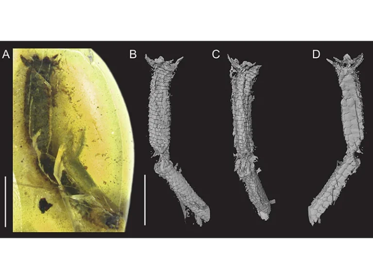

Hidden for decades inside a forgotten museum specimen drawer at the Steinhardt Museum of Natural History at Tel Aviv University, an extraordinary piece of Cretaceous Burmese amber dating back roughly 100 million years has yielded the world’s first direct, incontrovertible soft-tissue evidence of active caudal autotomy—the specialized predatory evasion behavior whereby lizards voluntarily sever their tails to escape capture. Utilizing advanced micro-computed tomography (micro-CT), an international team of researchers peered inside the clouding resin to uncover the pristine, uninjured muscle rosette and fracture plane identical to modern gecko escape mechanics.
A Serendipitous Discovery: Transforming a Fossilized Twig into a Lizard Tail
The discovery of this milestone fossil reads like an archaeological thriller. Sourced from the amber mines of northern Myanmar’s Hukawng Valley, the 18-millimeter resin nodule resided in the research archives of Tel Aviv University’s Steinhardt Museum of Natural History, designated under an unassuming catalog label identifying it as containing a minor botanical inclusion—likely a conifer twig or vegetative fragment.
That assumption evaporated when PhD researcher Dolev Fabrikant (Dolev Fabrikant) re-examined the specimen under high-magnification stereomicroscopy. Clearing the outer oxidized rind, Fabrikant recognized that the supposed plant surface was covered in finely overlapping, hexagonal epidermal scales characteristic of lacertilian reptiles.
Because internal fractures and organic turbidity obscured visual inspection, destructive physical preparation was out of the question. To evaluate the anatomy of the enigmatic inclusion while preserving its integrity, the team turned to non-invasive X-ray microtomography.
Micro-CT Reconstruction: Virtual Dissection of Cretaceous Soft Anatomy
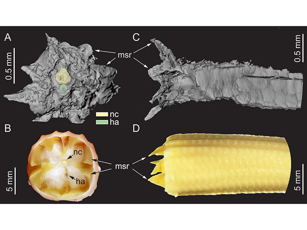
Employing micro-computed tomography with a spatial resolution under two micrometers, the researchers assembled a complete volumetric digital reconstruction of the inclusion, enabling virtual cross-sections through the ancient reptile’s tissues.
The scans revealed the distal tail section of a juvenile lizard that scurried across Gondwanan forest floors 100 million years ago. Crucially, the proximal detachment terminus exhibited no crushing, no lacerations, and no predator tooth puncture marks.
Instead, the separation surface displayed an impeccably symmetrical, wedge-shaped margin. The preserved myotomal bundles formed an exquisite interlocking zigzag configuration known as a muscle rosette—a structural signature that occurs exclusively when specialized tail detachment muscles undergo active contraction.
The tree resin fossilized during the mid-Cretaceous Cenomanian stage as dinosaurs dominated ancient terrestrial landscapes.
The tiny gemstone was cataloged for decades as an inconsequential plant shoot before microscopes revealed reptile scales.
Micro-CT 3D reconstruction confirmed the clean, zigzagging muscle plane unique to voluntary physiological detachment.
The earliest direct physical verification of specialized vascular and muscular sphincters operating in ancient vertebrates.
The Biophysics of Autotomy: An Evolutionary Masterpiece of Survival
Caudal autotomy represents one of the most sophisticated anti-predator adaptations in the animal kingdom. When a predator seizes a lizard by its tail, afferent neural pathways trigger an explosive, coordinated contraction of transverse caudal musculature.
Detachment occurs not between vertebrae, but directly across an intravertebral fracture plane traversing the centrum. Concurrently, arterial sphincters and venous valves instantly constrict, arresting blood flow and preventing exsanguination or infection at the stump.
Meanwhile, the severed tail continues to thrash and writhe violently for several minutes, driven by local glycogen reserves and autonomous spinal cord motor circuits. The thrashing tail captures the predator’s attention, granting the lizard critical seconds to dart into a secure rock fissure.
«Prior paleontological evidence was purely skeletal and inferential. This specimen captures the living biological act itself: the pristine, wedge-shaped rosette of muscle fibers confirms the tail was not bitten off or severed by trauma, but voluntarily shed 100 million years ago.»
Dolev Fabrikant (Dolev Fabrikant), Doctoral Researcher, Tel Aviv University
Paleontological Breakthrough: Bridging the Gap from Bone to Soft Tissue
Until this discovery, the evolutionary timeline of autotomy was pieced together through skeletal hints—such as fossil vertebrae showing cartilaginous regeneration pegs or transverse fracture grooves in extinct squamates.
However, skeletal architecture cannot confirm whether physiological muscular detachment was operational, or whether it was a dormant evolutionary precursor. Soft tissues rarely endure in traditional sedimentary rock formations, typically decaying into indistinct carbonaceous smears.
Just as advanced morphological studies illuminate ancient evolutionary transformations—such as recent paleobiological work showing how exceptional fossils decode the skeletal and feather transitions of Mesozoic flying reptiles—amber acts as a supreme preservation medium, capturing cellular-level biological behavior.
- Predetermined intravertebral fracture planes bifurcating individual caudal vertebrae;
- Specialized vascular sphincter muscles that instantly constrict caudal vessels to eliminate hemorrhage;
- Interlocking W-shaped muscle rosettes engineered to disengage without traumatic tissue laceration;
- Autonomous post-detachment spinal neuromuscular firing producing frantic tail thrashing to divert predator focus.
Cretaceous Predators: The Canopy Terrors of the Hukawng Ecosystem
The mid-Cretaceous forests of Myanmar were fraught with relentless predatory threats. The dense canopy was populated by feathered dromaeosaurs, enantiornithine birds, and giant predatory arthropods like scolopendrid centipedes and assassin bugs.
The juvenile lizard likely shed its tail during a life-or-death encounter with an avian hunter. Tumbling through the canopy, the thrashing tail lodged into a fresh seep of araucarian resin, which sealed the organic material from microbial decay.
The uncanny resemblance between this 100-million-year-old muscle rosette and detachment surfaces in living geckos and skinks underscores the extraordinary evolutionary resilience of autotomy, proving that its core biomechanics were perfected during the zenith of the dinosaur age.
The Renaissance of Museum Collections: Unlocking Forgotten Gems
The breakthrough spearheaded by Tel Aviv University highlights the immense scientific riches awaiting rediscovery inside long-established museum cabinets. Thousands of vintage amber and fossil specimens collected over past centuries were analyzed with basic optics and often misidentified.
With contemporary micro-CT and synchrotron imaging, researchers can achieve major evolutionary breakthroughs without setting foot in the field. This ancient lizard tail reminds us that history is not merely preserved in rocks, but continually waiting to be revealed through modern eyes.
Energetic Costs of Caudal Autotomy and Imperfect Regeneration
While self-amputation offers immediate salvation from predators, it exacts an enormous metabolic tax on the surviving reptile. In squamates, the caudal appendage functions as the primary adipose reserve, storing vital lipid depots crucial for surviving seasonal droughts and fueling vitellogenesis during reproduction. Losing the tail impairs locomotion speed, compromises aerial balance during leaps, and lowers social dominance hierarchies.
Furthermore, regenerating a secondary tail requires intensive energetic reallocation. The reconstructed tail lacks true segmented bony vertebrae, forming instead a solid cartilaginous rod devoid of predefined fracture planes, meaning secondary autotomy becomes far more difficult. The Cretaceous specimen demonstrates a pristine primary detachment, preserving the original musculoskeletal geometry in its purest evolutionary form.