

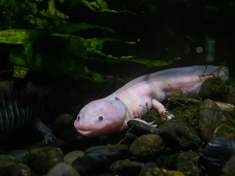

Dwelling in the high-altitude freshwater canals of Lake Xochimilco in Mexico is an enigmatic amphibian that directly challenges the conventional biological dogmas of senescence and tissue scarring. The axolotl regeneration blastema regenerative medicine empowers this creature to regenerate complex amputated limbs, retinal tissue, spinal cords, functioning myocardium, and even substantial portions of its cerebral cortex throughout its lifespan without forming fibrotic scar tissue. Decoding the colossal 32-gigabase genome of this neotenic wonder has unlocked fundamental developmental pathways that contemporary medicine is urgently working to translate into human regenerative therapeutics.
Neoteny and Eternal Youth: The Evolutionary Marvel of Ambystoma mexicanum
The axolotl represents a classic neotenic salamander belonging to the genus Ambystoma, endemic to the ancient volcanic lacustrine system of the Valley of Mexico. Unlike typical amphibians that transition through a radical metamorphic phase—shedding gills, developing lungs, and transitioning to a terrestrial existence—the axolotl remains permanently locked in its aquatic larval morphology. A congenital thyroid deficiency resulting from diminished pituitary stimulation leaves the animal with insufficient thyroxine levels to trigger metamorphosis.
Consequently, the axolotl retains juvenile anatomical hallmarks throughout its adult life: flamboyant external feathery gills, a broad dorsal caudal fin, a largely cartilaginous skeleton, and an aquatic metabolism, all while possessing fully functional reproductive organs. This state of physiological arrested development endows the axolotl with an unprecedented degree of cellular plasticity, an evolutionary capability largely silenced in adult mammals.
Revered in Aztec cosmology as the earthly avatar of Xolotl, the canine god of fire and transformation who disguised himself in the waterways to escape sacrifice, the axolotl has evolved into the quintessential laboratory model for unraveling the biological secrets of macroscopic limb and organ replacement.
The Mechanics of the Blastema: Cellular Dedifferentiation in Real Time
In adult mammals, traumatic tissue injury invariably elicits an emergency fibrotic response. Circulating fibroblasts converge on the wound bed, rapidly depositing disorganized, cross-linked type-I collagen fibers to seal the lesion. While this evolutionary adaptation prevents lethal hemorrhage and systemic sepsis, it creates a rigid mechanical barrier—the scar—that permanently precludes tissue morphogenesis and functional regeneration.
In stark contrast, the axolotl circumvents fibrosis through an exquisite molecular choreography. Within hours of limb amputation, specialized epidermal cells migrate across the severed stump, establishing a thickened signaling boundary known as the apical epithelial cap (AEC). The AEC immediately secretes an array of morphogenetic ligands, primarily fibroblast growth factors (FGF8, FGF2) and Wnt family proteins, instructing underlying tissues to commence dedifferentiation.
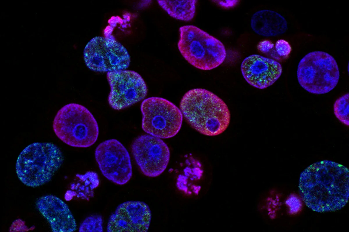
Under these signals, mature, post-mitotic somatic cells—including skeletal myocytes, dermal fibroblasts, chondrocytes, and Schwann cells—strip away their differentiated state. They re-enter the cell cycle as lineage-restricted multipotent progenitor cells, forming a specialized cone of proliferating tissue termed the blastema. Functioning as a high-fidelity analog of the embryonic limb bud, the blastema interprets positional cues along three spatial axes, faithfully rebuilding bone, muscle, vascular networks, and digits.
Following amputation, mature specialized cells (muscle, nerve, cartilage) shed differentiation markers, forming a proliferative stem-cell-like aggregate called the blastema.
Specialized regulatory macrophages suppress fibrotic extracellular collagen deposition, establishing an optimal microenvironment for true anatomical restoration.
The Ambystoma mexicanum genome is ten times larger than the human genome, dominated by vast non-coding intronic sequences and active retrotransposons.
Upon ablation of up to a third of the telencephalon, ependymoglial progenitors initiate neurogenesis, reconstructing functioning neural networks and synapses.
The Immunological Paradox: Macrophages Suppress Fibrosis
Historical biological paradigms assumed that robust regenerative capacity required an inactive or primitive immune system. However, groundbreaking investigations led by Dr. James Godwin at the MDI Biological Laboratory overturned this assumption by revealing that innate immune cells—specifically macrophages—are indispensable mediators of epimorphic regeneration.
When researchers systematically depleted endogenous macrophages in axolotls using clodronate liposomes, limb regeneration collapsed entirely. Rather than generating a proliferative blastema, the amputated stump developed classic fibrotic scar tissue identical to that seen in injured mammals. Crucially, when macrophages were permitted to repopulate the tissue, the animal regained its remarkable scar-free regenerative competency.
Axolotl macrophages exhibit a profoundly anti-inflammatory phenotype. Upon injury, they release substantial concentrations of interleukin-10 (IL-10) and TGF-beta3 while suppressing pro-fibrotic matrix metalloproteinases and pro-inflammatory signaling cascades. They act as precision cellular conductors, clearing apoptotic debris while instructing local progenitor pools to construct anatomically perfect structures rather than rigid scar tissue.
«The axolotl does not simply patch wounded tissue — it reawakens an embryonic developmental cascade within an adult organism, reverting mature cells into multipotent progenitors without oncogenic risk.»
Dr. Elly Tanaka, Senior Scientist, Research Institute of Molecular Pathology in Vienna (IMP)
The 32-Gigabase Genome: Cracking the Largest Sequenced Animal DNA
Assembling the complete genetic blueprint of the axolotl represented one of the most formidable bioinformatic milestones of the twenty-first century. Spanning an astonishing 32 billion base pairs—over ten times the length of the human haploid genome—the genome was finally assembled in 2018 by an international consortium led by the Max Planck Institute of Molecular Cell Biology and Genetics in Dresden using PacBio long-read sequencing.
Genomic architecture revealed that the colossal size of the axolotl genome does not stem from whole-genome duplications, but rather from an unprecedented expansion of non-coding intronic sequences and long interspersed nuclear elements (LINE retrotransposons). Average intron lengths in the axolotl exceed human counterparts by an order of magnitude, functioning as massive regulatory landscapes for epigenetic chromatin remodeling.
Comparative transcriptomics identified evolutionary novelties unique to salamanders, including the Kazaldc1 gene which governs structural cartilage formation, and specific microRNAs (such as miR-21) that orchestrate dedifferentiation dynamics. Remarkably, despite sustaining rapid cell division rates within the blastema, axolotls are virtually immune to neoplastic malignancies: tumor-suppressor pathways, mediated by p53 and retinoblastoma (Rb) proteins, exhibit exceptional regulatory stringency, completely shielding the organism from cancer.
- Neoteny allows the axolotl to reach sexual maturity while retaining its larval aquatic anatomy and external gills.
- An individual axolotl can regenerate the identical limb over one hundred times without structural degradation or tumor formation.
- Tissue grafts between individuals exhibit near-zero allograft rejection due to unique immunological tolerance mechanisms.
- Administration of exogenous thyroxine hormone can force metamorphosis into a terrestrial adult tiger salamander.
Myocardial and Telencephalon Regeneration: Blueprint for Human Therapies
The regenerative prowess of the axolotl extends far beyond appendages into vital organ systems. Following the surgical excision of up to 25% of the ventricular myocardium, the axolotl heart does not succumb to fatal heart failure or rigid scarring. Surviving cardiomyocytes temporarily disassemble their contractile sarcomeres, re-enter the mitotic cycle, proliferate, and reconstruct healthy, vascularized muscular walls within two months.
Even more extraordinary is the axolotl’s ability to regenerate its central nervous system. Following mechanical ablation of the dorsal telencephalon—equivalent to the human cerebral cortex—specialized ependymoglial cells lining the ventricular zones act as resident neural stem cells. These progenitors proliferate, migrate into the lesion cavity, and differentiate into mature, electrically active neurons that project functional axons across prior pathways, fully restoring lost behavioral reflexes.
For human clinical medicine, these discoveries offer an extraordinary translational roadmap. Pathologies such as post-myocardial infarction heart failure and traumatic spinal cord transections are driven by mammalian fibroblastic scarring. Because humans harbor largely orthologous genetic machinery, identifying the regulatory switches that unlock the axolotl’s regenerative cascades will enable synthetic biologics, specialized macrophage infusions, and RNA therapeutics capable of stimulating latent regenerative programs within our own tissues. Also read our related investigative feature on advancements in molecular genetics and the foundational architecture of the genome.