

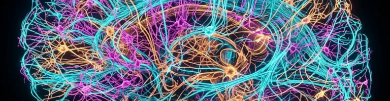

The international FlyWire Consortium has achieved one of the most monumental milestones in the history of biological science: researchers have unveiled the world’s first complete, wiring-level diagram (connectome) of an adult brain for a complex animal — the fruit fly *Drosophila melanogaster*. Spanning years of multidisciplinary collaboration between neuroscientists, computational physicists, and software engineers from Princeton University, the University of Cambridge, and HHMI’s Janelia Research Campus, the project successfully mapped all 139,255 neurons and charted over 54.5 million individual synapses. Published across a landmark collection of papers in the journal *Nature*, this digital atlas represents a long-awaited “Google Maps for the brain,” demystifying how neural circuits generate thought, sensory perception, and behavior.
From Worms to Complex Organisms: The Half-Century Quest of Connectomics
The ambitious dream of drafting an exhaustive wiring diagram of an animal nervous system originated in the 1970s, initiated by Nobel laureate Sydney Brenner in his pioneering reconstruction of the roundworm *Caenorhabditis elegans*. That heroic undertaking demanded more than twelve years of painstaking manual labor: the microscopic nematode possessed a modest 302 neurons linked by roughly 7,000 synapses, reconstructed slice-by-slice on transmission electron microscopy photographic prints.
Leaping from a simple soil roundworm to an adult fruit fly (*Drosophila melanogaster*) presented an exponential technological chasm. The brain of an adult fruit fly, though measuring less than one cubic millimeter, is hundreds of thousands of times more intricate: the fly executes breathtaking aerobatics, navigates by detecting polarized sunlight, evaluates courtship songs, recalls aversive odors, and modifies behavior through associative learning.
Overcoming this logistical barrier demanded profound innovations across multiple scientific disciplines: high-pressure tissue cryo-fixation, automated serial-section electron microscopy, and deep convolutional computer vision. A female fly brain was micro-sliced with a diamond knife into 7,050 contiguous sections merely 40 nanometers thick, generating over 100 terabytes of raw ultra-resolution imagery.
Unraveling these micro-architectures is equally foundational for the optimization of artificial neural networks and biomimetic robotics, where engineered systems seek to replicate biological computational efficiency.
Human-AI Symbiosis: How Global Crowdsourcing Built the FlyWire Map
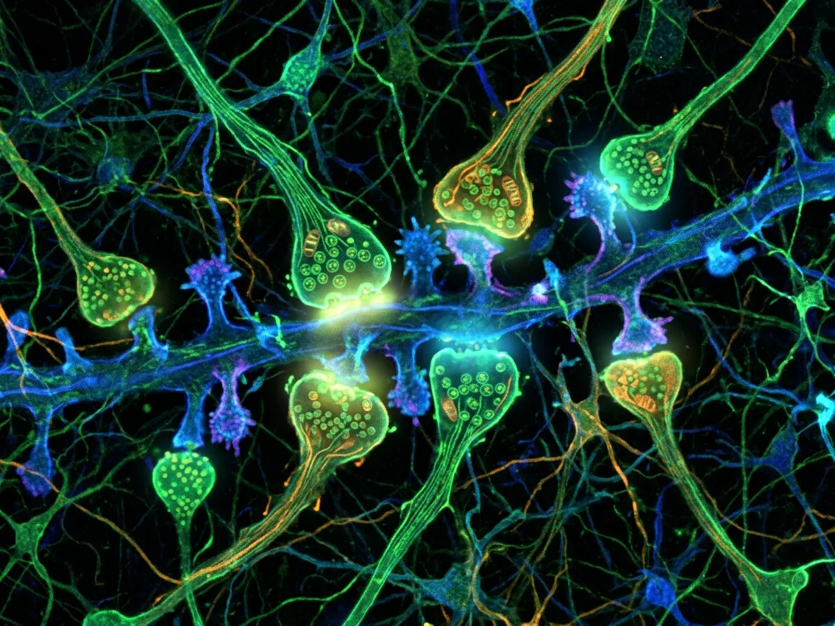
Had human researchers attempted to trace every individual neurite across seven thousand sections manually, the endeavor would have required centuries of uninterrupted effort. To surmount this bottleneck, researchers deployed deep convolutional neural networks trained to segment cellular boundaries and identify synaptic vesicles. However, even cutting-edge machine learning models suffered from segmentation errors, intermittently bridging adjacent membranes or truncating delicate dendritic spines.
The breakthrough lay in the architecture of the open-access FlyWire platform, which merged state-of-the-art computational vision with global crowdsourcing. The consortium mobilized hundreds of academic neurobiologists and passionate citizen scientists worldwide. Through an interactive 3D WebGL interface, proofreaders inspected flagged ambiguities, manually re-connecting broken branches and correcting algorithmic errors.
Through this monumental collective effort, the cumulative length of all reconstructed neuronal wiring reached an astonishing 150 meters — compressed inside a brain the size of a single poppy seed! Crucially, each reconstructed neuron was cross-referenced against neurotransmitter prediction models, classifying synapses as excitatory (cholinergic), inhibitory (GABAergic), or modulatory (glutamatergic and dopaminergic).
Researchers mapped every single nerve cell in the adult fly brain, cataloging over 4,000 previously unknown neuron types.
The digital connectome identifies every synaptic connection, classifying them into excitatory and inhibitory pathways.
The total cumulative length of microscopic axonal and dendritic fibers within the fly’s brain spans nearly 150 meters.
Machine learning performed initial segmentation, while citizen scientists and researchers proofread every circuit.
Anatomy of Thought: Steering Compasses and Visual Processing Hubs
The completed connectome uncovered an astounding structural organization within functional brain blocks. Researchers cataloged 8,453 distinct cell types, over four thousand of which had never before been described in biological literature. This underscores the staggering functional heterogeneity of living neural systems compared to the uniform layers characteristic of artificial neural networks.
A crown jewel of the connectome is the detailed mapping of the ellipsoid body within the central complex — the fly’s internal heading compass. Neurons in this circular neuropil are arranged in a ring attractor configuration: as the fly rotates, an active bump of electrical excitation shifts systematically around the ring, tracking spatial heading relative to celestial and visual cues in real time.
In the optic lobes, the connectome fully resolved the intricate circuits governing motion detection. Signals from thousands of individual ommatidia converge across laminar and medullary layers to perform split-second vector computations for obstacle avoidance. Remarkably, the fly’s brain executes these complex sensorimotor transformations while consuming merely microwatts of metabolic power.
From a developmental standpoint, the precise spatial steering of these axonal projections is governed by the universal principles of genetic transcription and cellular biology established during early morphogenesis.
«If we cannot fully understand how the brain of a tiny fruit fly functions, we have little hope of ever deciphering the human brain. The FlyWire connectome is not just a static circuit schematic; it is an active blueprint revealing how biological networks generate navigation, motor decisions, and memory.»
Dr. Mala Murthy, Director of the Princeton Neuroscience Institute
Computational Emulation: Running a Living Brain in Silico
The ultimate triumph of the connectome is that it transitions neuroscience from observational morphology into a rigorous predictive engineering discipline. Equipped with exact synaptic coordinates, directional polarities, and transmitter identities, scientists have begun simulating functional fly brain circuits entirely in silico.
In their flagship publication in Nature, the FlyWire team demonstrated a whole-brain computational model capable of simulating the neural pathways governing locomotion and walking cessation. When simulated sensory inputs were introduced, the in silico circuit accurately mirrored the physiological activations of a live insect, faithfully propagating signals down descending pathways to halt motor drive.
This establishes a historic paradigm shift: biologists can now test neuropharmacological interventions, genetic knockouts, and behavioral hypotheses inside high-fidelity digital twin simulations prior to performing in vivo experiments.
- The FlyWire Consortium findings were published in a dedicated special issue of Nature in October 2024.
- The specimen was microtomed into 7,050 sections of 40-nanometer thickness for serial transmission electron microscopy.
- The wiring diagram resolved the ring attractor circuit within the central complex that acts as a navigational compass.
- Computer simulations of the connectome successfully replicated walking cessation behaviors in silico.
The Bridge to the Human Brain: Unlocking Connectopathies and Medicine
Completing the Drosophila connectome establishes the algorithmic foundation for the next grand frontier: charting mammalian nervous systems. While the fruit fly possesses approximately 1.4×10^5 neurons, a mouse brain houses roughly 70 million neurons, and the human brain contains an astronomical 86 billion neurons intertwined by upwards of 100 trillion synapses.
Through the NIH BRAIN Initiative, international consortia are already scaling multibeam electron microscopy and exabyte-scale data pipelines. The crowdsourced, AI-assisted paradigm developed by FlyWire serves as the essential operational roadmap for handling these forthcoming data avalanches.
Beyond theoretical neurology, whole-brain connectomics holds revolutionary promise for clinical medicine. Devastating psychiatric and neurodegenerative conditions — including schizophrenia, autism spectrum disorders, and Alzheimer’s disease — are increasingly understood as “connectopathies”: structural breakdowns within the delicate topology of synaptic networks. Comprehensive connectomic maps will grant biomedical science the anatomical precision required to diagnose, model, and repair damaged neural circuitry.